DRT properties and production of x-rays. Basic properties of X-rays
X-ray radiation plays a huge role in modern medicine; the history of the discovery of X-rays dates back to the 19th century.
X-rays are electromagnetic waves that are produced with the participation of electrons. With strong acceleration of charged particles, artificial x-rays are created. It passes through special equipment:
- particle accelerators.
Discovery history
These rays were invented in 1895 by the German scientist Roentgen: while working with a cathode ray tube, he discovered the fluorescence effect of barium platinum cyanide. Then there was a description of such rays and their amazing ability to penetrate the tissues of the body. The rays began to be called x-rays (x-rays). Later in Russia they began to be called X-ray.
X-rays are able to penetrate even through walls. So Roentgen realized that he had made the greatest discovery in the field of medicine. It was from that time that separate sections in science began to form, such as radiology and radiology.
The rays are able to penetrate soft tissues, but are delayed, their length is determined by the obstacle of a hard surface. The soft tissues in the human body are the skin, and the hard tissues are the bones. In 1901, the scientist was awarded the Nobel Prize.
However, even before the discovery of Wilhelm Conrad Roentgen, other scientists were also interested in a similar topic. In 1853, the French physicist Antoine-Philiber Mason studied a high-voltage discharge between electrodes in a glass tube. The gas contained in it at low pressure began to emit a reddish glow. Pumping out excess gas from the tube led to the decay of the glow into a complex sequence of individual luminous layers, the hue of which depended on the amount of gas.
In 1878, William Crookes (English physicist) suggested that fluorescence occurs due to the impact of rays on the glass surface of the tube. But all these studies were not published anywhere, so Roentgen did not know about such discoveries. After the publication of his discoveries in 1895 in a scientific journal, where the scientist wrote that all bodies are transparent to these rays, albeit to a very different degree, other scientists became interested in similar experiments. They confirmed the invention of Roentgen, and further development and improvement of x-rays began.
Wilhelm Roentgen himself published two more scientific papers on the subject of x-rays in 1896 and 1897, after which he took up other activities. Thus, several scientists invented, but it was Roentgen who published scientific papers on this subject.

Imaging Principles
The features of this radiation are determined by the very nature of their appearance. Radiation occurs due to an electromagnetic wave. Its main properties include:
- Reflection. If the wave hits the surface perpendicularly, it will not be reflected. In some situations, a diamond has the property of reflection.
- The ability to penetrate tissue. In addition, the rays can pass through opaque surfaces of materials such as wood, paper, and the like.
- absorbency. Absorption depends on the density of the material: the denser it is, the more X-rays absorb it.
- Some substances fluoresce, that is, they glow. As soon as the radiation stops, the glow also disappears. If it continues after the cessation of the action of the rays, then this effect is called phosphorescence.
- X-rays can illuminate photographic film, just like visible light.
- If the beam passed through the air, then ionization occurs in the atmosphere. This state is called electrically conductive, and it is determined using a dosimeter, which sets the rate of radiation dosage.
Radiation - harm and benefit
When the discovery was made, the physicist Roentgen could not even imagine how dangerous his invention was. In the old days, all devices that produced radiation were far from perfect, and as a result, large doses of emitted rays were obtained. People did not understand the dangers of such radiation. Although some scientists even then put forward versions about the dangers of x-rays.

X-rays, penetrating into tissues, have a biological effect on them. The unit of measurement of radiation dose is roentgen per hour. The main influence is on the ionizing atoms that are inside the tissues. These rays act directly on the DNA structure of a living cell. The consequences of uncontrolled radiation include:
- cell mutation;
- the appearance of tumors;
- radiation burns;
- radiation sickness.
Contraindications for X-ray examinations:
- The patients are in critical condition.
- Pregnancy period due to negative effects on the fetus.
- Patients with bleeding or open pneumothorax.
How x-rays work and where it is used
- In medicine. X-ray diagnostics is used to translucent living tissues in order to identify certain disorders within the body. X-ray therapy is performed to eliminate tumor formations.
- In science. The structure of substances and the nature of X-rays are revealed. These issues are dealt with by such sciences as chemistry, biochemistry, crystallography.
- In industry. To detect violations in metal products.
- For the safety of the population. X-ray beams are installed at airports and other public places to scan luggage.

Medical use of X-ray radiation. X-rays are widely used in medicine and dentistry for the following purposes:
- For diagnosing diseases.
- For monitoring metabolic processes.
- For the treatment of many diseases.
The use of X-rays for medical purposes
In addition to detecting bone fractures, x-rays are widely used for medical purposes. The specialized application of x-rays is to achieve the following goals:
- To destroy cancer cells.
- To reduce the size of the tumor.
- To reduce pain.
For example, radioactive iodine, used in endocrinological diseases, is actively used in thyroid cancer, thereby helping many people get rid of this terrible disease. Currently, to diagnose complex diseases, X-rays are connected to computers, as a result, the latest research methods appear, such as computed axial tomography.
Such a scan provides doctors with color images that show the internal organs of a person. To detect the work of internal organs, a small dose of radiation is sufficient. X-rays are also widely used in physiotherapy.

Basic properties of X-rays
- penetrating ability. All bodies are transparent to the x-ray, and the degree of transparency depends on the thickness of the body. It is due to this property that the beam began to be used in medicine to detect the functioning of organs, the presence of fractures and foreign bodies in the body.
- They are able to cause the glow of some objects. For example, if barium and platinum are applied to cardboard, then, after passing through the beam scanning, it will glow greenish-yellow. If you place your hand between the X-ray tube and the screen, then the light will penetrate more into the bone than into the tissue, so the bone tissue will shine brightest on the screen, and the muscle tissue will be less bright.
- Action on film. X-rays can, like light, darken film, which makes it possible to photograph the shadow side that is obtained when objects are examined by x-rays.
- X-rays can ionize gases. This makes it possible not only to find rays, but also to reveal their intensity by measuring the ionization current in the gas.
- They have a biochemical effect on the body of living beings. Thanks to this property, X-rays have found their wide application in medicine: they can treat both skin diseases and diseases of internal organs. In this case, the desired dosage of radiation and the duration of the rays are selected. Prolonged and excessive use of such treatment is very harmful and detrimental to the body.
The consequence of the use of X-rays was the saving of many human lives. X-ray helps not only to diagnose the disease in a timely manner, treatment methods using radiation therapy relieve patients of various pathologies, from hyperfunction of the thyroid gland to malignant tumors of bone tissues.
Ministry of Education and Science of the Russian Federation
Federal Agency for Education
GOU VPO SUSU
Department of Physical Chemistry
at the KSE course: “X-ray radiation”
Completed:
Naumova Daria Gennadievna
Checked:
Associate Professor, K.T.N.
Tanklevskaya N.M.
Chelyabinsk 2010
Introduction
Chapter I. Discovery of X-rays
Receipt
Interaction with matter
Biological impact
registration
Application
How an x-ray is taken
natural x-rays
Chapter II. Radiography
Application
Image Acquisition Method
Benefits of radiography
Disadvantages of radiography
Fluoroscopy
Receipt principle
Benefits of Fluoroscopy
Disadvantages of Fluoroscopy
Digital technologies in fluoroscopy
Multiline scanning method
Conclusion
List of used literature
Introduction
X-ray radiation - electromagnetic waves, the photon energy of which is determined by the energy range from ultraviolet to gamma radiation, which corresponds to the wavelength range from 10−4 to 10² Å (from 10−14 to 10−8 m).
Like visible light, X-rays cause blackening of photographic film. This property is of great importance for medicine, industry and scientific research. Passing through the object under study and then falling on the film, X-ray radiation depicts its internal structure on it. Since the penetrating power of X-ray radiation is different for different materials, parts of the object that are less transparent to it give brighter areas in the photograph than those through which the radiation penetrates well. Thus, bone tissues are less transparent to x-rays than the tissues that make up the skin and internal organs. Therefore, on the radiograph, the bones will be indicated as lighter areas and the fracture site, which is more transparent for radiation, can be quite easily detected. X-ray imaging is also used in dentistry to detect caries and abscesses in the roots of teeth, as well as in industry to detect cracks in castings, plastics and rubbers.
X-rays are used in chemistry to analyze compounds and in physics to study the structure of crystals. An X-ray beam passing through a chemical compound causes a characteristic secondary radiation, the spectroscopic analysis of which allows the chemist to determine the composition of the compound. When falling on a crystalline substance, an X-ray beam is scattered by the atoms of the crystal, giving a clear, regular pattern of spots and stripes on a photographic plate, which makes it possible to establish the internal structure of the crystal.
The use of X-rays in cancer treatment is based on the fact that it kills cancer cells. However, it can also have an undesirable effect on normal cells. Therefore, extreme caution must be exercised in this use of X-rays.
Chapter I. Discovery of X-rays
The discovery of X-rays is attributed to Wilhelm Conrad Roentgen. He was the first to publish an article on X-rays, which he called x-rays (x-ray). An article by Roentgen titled "On a new type of rays" was published on December 28, 1895 in the journal of the Würzburg Physico-Medical Society. It is considered, however, proven that X-rays have already been obtained before. The cathode ray tube that Roentgen used in his experiments was developed by J. Hittorf and W. Crookes. This tube produces X-rays. This was shown in the experiments of Crookes and from 1892 in the experiments of Heinrich Hertz and his student Philipp Lenard through the blackening of photographic plates. However, none of them realized the significance of their discovery and did not publish their results. Also, Nikola Tesla, starting in 1897, experimented with cathode ray tubes, received X-rays, but did not publish his results.
For this reason, Roentgen did not know about the discoveries made before him and discovered the rays, later named after him, independently - while observing the fluorescence that occurs during the operation of a cathode ray tube. Roentgen studied X-rays for a little over a year (from November 8, 1895 to March 1897) and published only three relatively small articles about them, but they provided such a comprehensive description of the new rays that hundreds of papers by his followers, then published over the course of 12 years, could neither add nor change anything significant. Roentgen, who had lost interest in X-rays, told his colleagues: "I already wrote everything, don't waste your time." Also contributing to Roentgen's fame was the famous photograph of his wife's hand, which he published in his article (see image on the right). Such fame brought Roentgen in 1901 the first Nobel Prize in Physics, and the Nobel Committee emphasized the practical importance of his discovery. In 1896, the name "X-rays" was first used. In some countries, the old name remains - X-rays. In Russia, the rays began to be called "X-ray" at the suggestion of a student V.K. Roentgen - Abram Fedorovich Ioffe.
Position on the scale of electromagnetic waves
The energy ranges of X-rays and gamma-rays overlap in a wide energy range. Both types of radiation are electromagnetic radiation and are equivalent for the same photon energy. The terminological difference lies in the mode of occurrence - X-rays are emitted with the participation of electrons (either in atoms or free ones), while gamma radiation is emitted in the processes of de-excitation of atomic nuclei. X-ray photons have energies from 100 eV to 250 keV, which corresponds to radiation with a frequency of 3 1016 Hz to 6 1019 Hz and a wavelength of 0.005 - 10 nm (there is no generally accepted definition of the lower limit of the X-ray range in the wavelength scale). Soft X-rays are characterized by the lowest photon energy and radiation frequency (and the longest wavelength), while hard X-rays have the highest photon energy and radiation frequency (and the shortest wavelength).
(X-ray photograph (roentgenogram) of his wife's hand, taken by V.K. Roentgen)
)Receipt
X-rays are produced by strong acceleration of charged particles (mainly electrons) or by high-energy transitions in the electron shells of atoms or molecules. Both effects are used in X-ray tubes, in which electrons emitted from a hot cathode are accelerated (no X-rays are emitted, because the acceleration is too low) and hit the anode, where they are sharply decelerated (X-rays are emitted: the so-called . bremsstrahlung) and at the same time knock out electrons from the inner electron shells of the atoms of the metal from which the anode is made. Empty spaces in the shells are occupied by other electrons of the atom. In this case, X-ray radiation is emitted with a certain energy characteristic of the anode material (characteristic radiation, frequencies are determined by the Moseley law:
,where Z is the atomic number of the anode element, A and B are constants for a certain value of the principal quantum number n of the electron shell). At present, anodes are made mainly of ceramics, and the part where the electrons hit is made of molybdenum. In the process of acceleration-deceleration, only 1% of the kinetic energy of the electron goes to X-rays, 99% of the energy is converted into heat.
X-rays can also be obtained in particle accelerators. so-called. synchrotron radiation occurs when a beam of particles is deflected in a magnetic field, as a result of which they experience acceleration in a direction perpendicular to their motion. Synchrotron radiation has a continuous spectrum with an upper limit. With appropriately chosen parameters (the magnitude of the magnetic field and the energy of the particles), X-rays can also be obtained in the spectrum of synchrotron radiation.

Schematic representation of an x-ray tube. X - x-rays, K - cathode, A - anode (sometimes called anticathode), C - heat sink, Uh - cathode filament voltage, Ua - accelerating voltage, Win - water cooling inlet, Wout - water cooling outlet (see x-ray tube) .
Interaction with matter
The refractive index of almost any substance for x-rays differs little from unity. A consequence of this is the fact that there is no material from which an X-ray lens could be made. In addition, when X-rays are incident perpendicular to the surface, they are almost not reflected. Despite this, in X-ray optics, methods have been found for constructing optical elements for X-rays.
X-rays can penetrate matter, and different substances absorb them differently. The absorption of x-rays is their most important property in x-ray photography. The intensity of X-rays decreases exponentially depending on the path traveled in the absorbing layer (I = I0e-kd, where d is the layer thickness, the coefficient k is proportional to Z3λ3, Z is the atomic number of the element, λ is the wavelength).
Absorption occurs as a result of photoabsorption and Compton scattering:
Photoabsorption is understood as the process of knocking out an electron from the shell of an atom by a photon, which requires that the photon energy be greater than a certain minimum value. If we consider the probability of the act of absorption depending on the energy of the photon, then when a certain energy is reached, it (probability) increases sharply to its maximum value. For higher energies, the probability continuously decreases. Because of this dependence, it is said that there is an absorption limit. The place of the electron knocked out during the act of absorption is occupied by another electron, while radiation with a lower photon energy is emitted, the so-called. fluorescence process.
X-rays, invisible radiation capable of penetrating, albeit to varying degrees, all substances. It is electromagnetic radiation with a wavelength of about 10-8 cm.
Like visible light, X-rays cause blackening of photographic film. This property is of great importance for medicine, industry and scientific research. Passing through the object under study and then falling on the film, X-ray radiation depicts its internal structure on it. Since the penetrating power of X-ray radiation is different for different materials, parts of the object that are less transparent to it give brighter areas in the photograph than those through which the radiation penetrates well. Thus, bone tissues are less transparent to x-rays than the tissues that make up the skin and internal organs. Therefore, on the radiograph, the bones will be indicated as lighter areas and the fracture site, which is more transparent for radiation, can be quite easily detected. X-ray imaging is also used in dentistry to detect caries and abscesses in the roots of teeth, as well as in industry to detect cracks in castings, plastics and rubbers.
X-rays are used in chemistry to analyze compounds and in physics to study the structure of crystals. An X-ray beam passing through a chemical compound causes a characteristic secondary radiation, the spectroscopic analysis of which allows the chemist to determine the composition of the compound. When falling on a crystalline substance, an X-ray beam is scattered by the atoms of the crystal, giving a clear, regular pattern of spots and stripes on a photographic plate, which makes it possible to establish the internal structure of the crystal.
The use of X-rays in cancer treatment is based on the fact that it kills cancer cells. However, it can also have an undesirable effect on normal cells. Therefore, extreme caution must be exercised in this use of X-rays.
Getting x-rays
X-ray radiation occurs when electrons moving at high speeds interact with matter. When electrons collide with atoms of any substance, they quickly lose their kinetic energy. In this case, most of it is converted into heat, and a small fraction, usually less than 1%, is converted into X-ray energy. This energy is released in the form of quanta - particles called photons that have energy but have zero rest mass. X-ray photons differ in their energy, which is inversely proportional to their wavelength. With the usual method of obtaining X-rays, a wide range of wavelengths is obtained, which is called the X-ray spectrum.
X-ray tubes. In order to obtain X-ray radiation due to the interaction of electrons with matter, it is necessary to have a source of electrons, means of accelerating them to high speeds, and a target capable of withstanding electron bombardment and producing X-ray radiation of the desired intensity. The device that has all this is called an x-ray tube. Early explorers used "deep vacuum" tubes such as today's discharge tubes. The vacuum in them was not very high.
Discharge tubes contain a small amount of gas, and when a large potential difference is applied to the electrodes of the tube, the gas atoms turn into positive and negative ions. The positive ones move towards the negative electrode (cathode) and, falling on it, knock electrons out of it, and they, in turn, move towards the positive electrode (anode) and, bombarding it, create a stream of X-ray photons.
In the modern X-ray tube developed by Coolidge (Fig. 11), the source of electrons is a tungsten cathode heated to a high temperature.
Rice. eleven.
The electrons are accelerated to high speeds by the high potential difference between the anode (or anticathode) and the cathode. Since the electrons must reach the anode without colliding with atoms, a very high vacuum is required, for which the tube must be well evacuated. This also reduces the probability of ionization of the remaining gas atoms and the associated side currents.
When bombarded with electrons, the tungsten anticathode emits characteristic x-rays. The cross section of the X-ray beam is less than the actual irradiated area. 1 - electron beam; 2 - cathode with a focusing electrode; 3 - glass shell (tube); 4 - tungsten target (anticathode); 5 - cathode filament; 6 - actually irradiated area; 7 - effective focal spot; 8 - copper anode; 9 - window; 10 - scattered x-rays.
The electrons are focused on the anode by a specially shaped electrode surrounding the cathode. This electrode is called the focusing electrode and, together with the cathode, forms the "electronic spotlight" of the tube. The anode subjected to electron bombardment must be made of a refractory material, since most of the kinetic energy of the bombarding electrons is converted into heat. In addition, it is desirable that the anode be made of a material with a high atomic number, since the x-ray yield increases with increasing atomic number. Tungsten, whose atomic number is 74, is most often chosen as the anode material. The design of X-ray tubes can be different depending on the application conditions and requirements.
Radiology is a section of radiology that studies the effects of X-ray radiation on the body of animals and humans arising from this disease, their treatment and prevention, as well as methods for diagnosing various pathologies using X-rays (X-ray diagnostics). A typical X-ray diagnostic apparatus includes a power supply (transformers), a high-voltage rectifier that converts the alternating current of the electrical network into direct current, a control panel, a tripod and an X-ray tube.
X-rays are a type of electromagnetic oscillations that are formed in an X-ray tube during a sharp deceleration of accelerated electrons at the moment of their collision with the atoms of the anode substance. At present, the point of view is generally accepted that X-rays, by their physical nature, are one of the types of radiant energy, the spectrum of which also includes radio waves, infrared rays, visible light, ultraviolet rays and gamma rays of radioactive elements. X-ray radiation can be characterized as a collection of its smallest particles - quanta or photons.
Rice. 1 - mobile x-ray machine:
A - x-ray tube;
B - power supply;
B - adjustable tripod.
 Rice. 2 - X-ray machine control panel (mechanical - on the left and electronic - on the right):
 A - panel for adjusting exposure and hardness;
B - high voltage supply button.
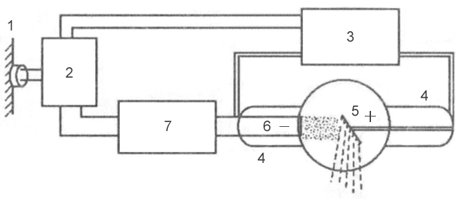 Rice. 3 is a block diagram of a typical x-ray machine
 1 - network;
2 - autotransformer;
3 - step-up transformer;
4 - x-ray tube;
5 - anode;
6 - cathode;
7 - step-down transformer.
Mechanism of X-ray generation
X-rays are formed at the moment of collision of a stream of accelerated electrons with the anode material. When electrons interact with a target, 99% of their kinetic energy is converted into thermal energy and only 1% into X-rays.
An X-ray tube consists of a glass container in which 2 electrodes are soldered: a cathode and an anode. Air is pumped out of the glass cylinder: the movement of electrons from the cathode to the anode is possible only under conditions of relative vacuum (10 -7 -10 -8 mm Hg). On the cathode there is a filament, which is a tightly twisted tungsten filament. When an electric current is applied to the filament, electron emission occurs, in which electrons are separated from the spiral and form an electron cloud near the cathode. This cloud is concentrated at the focusing cup of the cathode, which sets the direction of electron movement. Cup - a small depression in the cathode. The anode, in turn, contains a tungsten metal plate on which the electrons are focused - this is the site of the formation of x-rays.
Rice. 4 - X-ray tube device: A - cathode; 
B - anode;
B - tungsten filament;
G - focusing cup of the cathode;
D - stream of accelerated electrons;
E - tungsten target;
G - glass flask;
З - a window from beryllium;
And - formed x-rays;
K - aluminum filter.
2 transformers are connected to the electron tube: step-down and step-up. A step-down transformer heats the tungsten filament with a low voltage (5-15 volts), resulting in electron emission. A step-up, or high-voltage, transformer goes directly to the cathode and anode, which are supplied with a voltage of 20–140 kilovolts. Both transformers are placed in the high-voltage block of the X-ray machine, which is filled with transformer oil, which provides cooling of the transformers and their reliable insulation.
After an electron cloud has formed with the help of a step-down transformer, the step-up transformer is turned on, and high-voltage voltage is applied to both poles of the electrical circuit: a positive pulse to the anode, and a negative pulse to the cathode. Negatively charged electrons are repelled from a negatively charged cathode and tend to a positively charged anode - due to such a potential difference, a high speed of movement is achieved - 100 thousand km / s. At this speed, electrons bombard the tungsten anode plate, completing an electrical circuit, resulting in X-rays and thermal energy.
X-ray radiation is subdivided into bremsstrahlung and characteristic. Bremsstrahlung occurs due to a sharp deceleration of the speed of electrons emitted by a tungsten filament. Characteristic radiation occurs at the moment of rearrangement of the electron shells of atoms. Both of these types are formed in an X-ray tube at the moment of collision of accelerated electrons with atoms of the anode material. The emission spectrum of an X-ray tube is a superposition of bremsstrahlung and characteristic X-rays.
 Rice. 5 - the principle of the formation of bremsstrahlung X-rays.
 Rice. 6 - the principle of formation of the characteristic x-rays.
Basic properties of X-rays
- X-rays are invisible to visual perception.
- X-ray radiation has a great penetrating power through the organs and tissues of a living organism, as well as dense structures of inanimate nature, which do not transmit visible light rays.
- X-rays cause certain chemical compounds to glow, called fluorescence.
- Zinc and cadmium sulfides fluoresce yellow-green,
- Crystals of calcium tungstate - violet-blue.
Scale of electromagnetic oscillations
X-rays have a specific wavelength and frequency of oscillation. Wavelength (λ) and oscillation frequency (ν) are related by the relationship: λ ν = c, where c is the speed of light, rounded to 300,000 km per second. The energy of X-rays is determined by the formula E = h ν, where h is Planck's constant, a universal constant equal to 6.626 10 -34 J⋅s. The wavelength of the rays (λ) is related to their energy (E) by the relation: λ = 12.4 / E.
X-ray radiation differs from other types of electromagnetic oscillations in wavelength (see table) and quantum energy. The shorter the wavelength, the higher its frequency, energy and penetrating power. The X-ray wavelength is in the range
. By changing the wavelength of X-ray radiation, it is possible to control its penetrating power. X-rays have a very short wavelength, but a high frequency of oscillation, so they are invisible to the human eye. Due to their enormous energy, quanta have a high penetrating power, which is one of the main properties that ensure the use of X-rays in medicine and other sciences.X-ray characteristics
Intensity- quantitative characteristic of x-ray radiation, which is expressed by the number of rays emitted by the tube per unit time. The intensity of X-rays is measured in milliamps. Comparing it with the intensity of visible light from a conventional incandescent lamp, we can draw an analogy: for example, a 20-watt lamp will shine with one intensity, or power, and a 200-watt lamp will shine with another, while the quality of the light itself (its spectrum) is the same . The intensity of X-ray radiation is, in fact, its quantity. Each electron creates one or more radiation quanta on the anode, therefore, the amount of X-rays during exposure of the object is regulated by changing the number of electrons tending to the anode and the number of interactions of electrons with atoms of the tungsten target, which can be done in two ways:
- By changing the degree of incandescence of the cathode spiral using a step-down transformer (the number of electrons generated during emission will depend on how hot the tungsten spiral is, and the number of radiation quanta will depend on the number of electrons);
- By changing the value of the high voltage supplied by the step-up transformer to the poles of the tube - the cathode and the anode (the higher the voltage is applied to the poles of the tube, the more kinetic energy the electrons receive, which, due to their energy, can interact with several atoms of the anode substance in turn - see Fig. rice. five; electrons with low energy will be able to enter into a smaller number of interactions).
The X-ray intensity (anode current) multiplied by the shutter speed (tube time) corresponds to the X-ray exposure, which is measured in mAs (milliamps per second). Exposure is a parameter that, like intensity, characterizes the amount of rays emitted by an x-ray tube. The only difference is that the exposure also takes into account the operating time of the tube (for example, if the tube works for 0.01 sec, then the number of rays will be one, and if 0.02 sec, then the number of rays will be different - twice more). The radiation exposure is set by the radiologist on the control panel of the X-ray machine, depending on the type of study, the size of the object under study and the diagnostic task.
Rigidity- qualitative characteristic of x-ray radiation. It is measured by the high voltage on the tube - in kilovolts. Determines the penetrating power of x-rays. It is regulated by the high voltage supplied to the X-ray tube by a step-up transformer. The higher the potential difference is created on the electrodes of the tube, the more force the electrons repel from the cathode and rush to the anode, and the stronger their collision with the anode. The stronger their collision, the shorter the wavelength of the resulting X-ray radiation and the higher the penetrating power of this wave (or the hardness of the radiation, which, like the intensity, is regulated on the control panel by the voltage parameter on the tube - kilovoltage).
λ - wavelength;  Rice. 7 - Dependence of the wavelength on the energy of the wave:
E - wave energy  Rice. 8 - The ratio of the voltage on the X-ray tube and the wavelength of the resulting X-ray radiation:
Classification of x-ray tubes
- By appointment
- Diagnostic
- Therapeutic
- For structural analysis
- For transillumination
- By design
- By focus
- Single-focus (one spiral on the cathode, and one focal spot on the anode)
- Bifocal (two spirals of different sizes on the cathode, and two focal spots on the anode)
- By type of anode
- Stationary (fixed)
- Rotating
X-rays are used not only for radiodiagnostic purposes, but also for therapeutic purposes. As noted above, the ability of X-ray radiation to suppress the growth of tumor cells makes it possible to use it in radiation therapy of oncological diseases. In addition to the medical field of application, X-ray radiation has found wide application in the engineering and technical field, materials science, crystallography, chemistry and biochemistry: for example, it is possible to identify structural defects in various products (rails, welds, etc.) using X-ray radiation. The type of such research is called defectoscopy. And at airports, railway stations and other crowded places, X-ray television introscopes are actively used to scan hand luggage and luggage for security purposes.
Depending on the type of anode, X-ray tubes differ in design. Due to the fact that 99% of the kinetic energy of the electrons is converted into thermal energy, during the operation of the tube, the anode is significantly heated - the sensitive tungsten target often burns out. The anode is cooled in modern x-ray tubes by rotating it. The rotating anode has the shape of a disk, which distributes heat evenly over its entire surface, preventing local overheating of the tungsten target.
The design of X-ray tubes also differs in focus. Focal spot - the section of the anode on which the working X-ray beam is generated. It is subdivided into the real focal spot and the effective focal spot ( rice. 12). Due to the angle of the anode, the effective focal spot is smaller than the real one. Different focal spot sizes are used depending on the size of the image area. The larger the image area, the wider the focal spot must be to cover the entire image area. However, a smaller focal spot produces better image clarity. Therefore, when producing small images, a short filament is used and the electrons are directed to a small area of the anode target, creating a smaller focal spot.
 Rice. 9 - x-ray tube with a stationary anode.
 Rice. 10 - X-ray tube with a rotating anode.
 Rice. 11 - X-ray tube device with a rotating anode.
 Rice. 12 is a diagram of the formation of a real and effective focal spot.
Modern medical diagnostics and treatment of certain diseases cannot be imagined without devices that use the properties of X-rays. The discovery of X-rays occurred more than 100 years ago, but even now work continues on the creation of new methods and apparatus to minimize the negative effect of radiation on the human body.
Who and how discovered X-rays
Under natural conditions, the flux of X-rays is rare and is emitted only by certain radioactive isotopes. X-rays or X-rays were only discovered in 1895 by the German scientist Wilhelm Röntgen. This discovery happened by chance, during an experiment to study the behavior of light rays under conditions approaching vacuum. The experiment involved a cathode gas discharge tube with reduced pressure and a fluorescent screen, which each time began to glow at the moment when the tube began to act.
Intrigued by the strange effect, Roentgen conducted a series of studies showing that the resulting radiation, invisible to the eye, can penetrate various obstacles: paper, wood, glass, some metals, and even through the human body. Despite the lack of understanding of the very nature of what is happening, whether such a phenomenon is caused by the generation of a stream of unknown particles or waves, the following pattern was noted - radiation easily passes through the soft tissues of the body, and much harder through solid living tissues and inanimate substances.
Roentgen was not the first to study this phenomenon. In the middle of the 19th century, Frenchman Antoine Mason and Englishman William Crookes studied similar possibilities. However, it was Roentgen who first invented the cathode tube and an indicator that could be used in medicine. He was the first to publish a scientific work, which brought him the title of the first Nobel laureate among physicists.
In 1901, a fruitful collaboration began between the three scientists, who became the founding fathers of radiology and radiology.

X-ray properties
X-rays are an integral part of the general spectrum of electromagnetic radiation. The wavelength is between gamma and ultraviolet rays. X-rays have all the usual wave properties:
- diffraction;
- refraction;
- interference;
- propagation speed (it is equal to light).
To artificially generate an X-ray flux, special devices are used - X-ray tubes. X-ray radiation arises from the contact of fast tungsten electrons with substances evaporating from a hot anode. Against the background of interaction, short-length electromagnetic waves arise, which are in the spectrum from 100 to 0.01 nm and in the energy range of 100-0.1 MeV. If the wavelength of the rays is less than 0.2 nm - this is hard radiation, if the wavelength is greater than the specified value, they are called soft x-rays.
It is significant that the kinetic energy arising from the contact of electrons and the anode substance is 99% converted into heat energy and only 1% is X-rays.
X-ray radiation - bremsstrahlung and characteristic
X-radiation is a superposition of two types of rays - bremsstrahlung and characteristic. They are generated in the handset simultaneously. Therefore, X-ray irradiation and the characteristic of each specific X-ray tube - the spectrum of its radiation, depends on these indicators, and represents their superposition.
Bremsstrahlung or continuous X-rays are the result of deceleration of electrons evaporating from a tungsten filament.
Characteristic or line X-rays are formed at the moment of rearrangement of the atoms of the substance of the anode of the X-ray tube. The wavelength of the characteristic rays directly depends on the atomic number of the chemical element used to make the anode of the tube.
The listed properties of X-rays allow them to be used in practice:
- invisible to the ordinary eye;
- high penetrating ability through living tissues and inanimate materials that do not transmit visible light;
- ionization effect on molecular structures.
Principles of X-ray Imaging
The property of x-rays on which imaging is based is the ability to either decompose or cause some substances to glow.
X-ray irradiation causes a fluorescent glow in cadmium and zinc sulfides - green, and in calcium tungstate - blue. This property is used in the technique of medical X-ray transillumination, and also increases the functionality of X-ray screens.
The photochemical effect of X-rays on light-sensitive silver halide materials (illumination) makes it possible to carry out diagnostics - to take X-ray images. This property is also used in measuring the amount of the total dose that laboratory assistants receive in X-ray rooms. Wearable dosimeters have special sensitive tapes and indicators. The ionizing effect of X-ray radiation makes it possible to determine the qualitative characteristics of the obtained X-rays.
A single exposure to conventional X-rays increases the risk of cancer by only 0.001%.
Areas where X-rays are used
The use of X-rays is acceptable in the following industries:
- Security. Fixed and portable devices for detecting dangerous and prohibited items at airports, customs or in crowded places.
- Chemical industry, metallurgy, archeology, architecture, construction, restoration work - to detect defects and conduct chemical analysis of substances.
- Astronomy. It helps to observe cosmic bodies and phenomena with the help of X-ray telescopes.
- military industry. For the development of laser weapons.
The main application of X-rays is in the medical field. Today, the section of medical radiology includes: radiodiagnostics, radiotherapy (X-ray therapy), radiosurgery. Medical universities produce highly specialized specialists - radiologists.
X-Radiation - harm and benefit, effects on the body
The high penetrating power and ionizing effect of X-rays can cause a change in the structure of the DNA of the cell, therefore it is dangerous for humans. The harm from X-ray radiation is directly proportional to the received radiation dose. Different organs respond to irradiation to varying degrees. The most susceptible include:
- bone marrow and bone tissue;
- lens of the eye;
- thyroid;
- mammary and sex glands;
- lung tissue.
Uncontrolled use of X-ray radiation can cause reversible and irreversible pathologies.
Consequences of X-ray exposure:
- damage to the bone marrow and the occurrence of pathologies of the hematopoietic system - erythrocytopenia, thrombocytopenia, leukemia;
- damage to the lens, with the subsequent development of cataracts;
- cellular mutations that are inherited;
- development of oncological diseases;
- getting radiation burns;
- development of radiation sickness.
Important! Unlike radioactive substances, X-rays do not accumulate in the tissues of the body, which means that there is no need to remove X-rays from the body. The harmful effect of X-rays ends when the medical device is turned off.
The use of X-rays in medicine is permissible not only for diagnostic (traumatology, dentistry), but also for therapeutic purposes:
- from x-rays in small doses, the metabolism in living cells and tissues is stimulated;
- certain limiting doses are used for the treatment of oncological and benign neoplasms.

Methods for diagnosing pathologies using X-rays
Radiodiagnostics includes the following methods:
- Fluoroscopy is a study in which an image is obtained on a fluorescent screen in real time. Along with the classical real-time imaging of a body part, today there are X-ray television transillumination technologies - the image is transferred from a fluorescent screen to a television monitor located in another room. Several digital methods have been developed for processing the resulting image, followed by transferring it from the screen to paper.
- Fluorography is the cheapest method for examining the chest organs, which consists in making a small picture of 7x7 cm. Despite the possibility of error, it is the only way to conduct a mass annual examination of the population. The method is not dangerous and does not require the withdrawal of the received radiation dose from the body.
- Radiography - obtaining a summary image on film or paper to clarify the shape of an organ, its position or tone. Can be used to assess peristalsis and the condition of the mucous membranes. If there is a choice, then among modern X-ray devices, preference should be given neither to digital devices, where the x-ray flux can be higher than that of old devices, but to low-dose X-ray devices with direct flat semiconductor detectors. They allow you to reduce the load on the body by 4 times.
- Computed X-ray tomography is a technique that uses x-rays to obtain the required number of images of sections of a selected organ. Among the many varieties of modern CT devices, low-dose high-resolution CT scanners are used for a series of repeated studies.

Radiotherapy
X-ray therapy refers to local treatment methods. Most often, the method is used to destroy cancer cells. Since the effect of exposure is comparable to surgical removal, this treatment method is often called radiosurgery.
Today, x-ray treatment is carried out in the following ways:
- External (proton therapy) - the radiation beam enters the patient's body from the outside.
- Internal (brachytherapy) - the use of radioactive capsules by implanting them into the body, with the placement closer to the cancerous tumor. The disadvantage of this method of treatment is that until the capsule is removed from the body, the patient needs to be isolated.
These methods are gentle, and their use is preferable to chemotherapy in some cases. Such popularity is due to the fact that the rays do not accumulate and do not require removal from the body, they have a selective effect, without affecting other cells and tissues.
Safe X-ray exposure rate
This indicator of the norm of permissible annual exposure has its own name - a genetically significant equivalent dose (GED). There are no clear quantitative values for this indicator.
- This indicator depends on the age and desire of the patient to have children in the future.
- It depends on which organs were examined or treated.
- The GZD is affected by the level of natural radioactive background of the region where a person lives.
Today, the following average GZD standards are in effect:
- the level of exposure from all sources, with the exception of medical ones, and without taking into account the natural radiation background - 167 mRem per year;
- the norm for an annual medical examination is not more than 100 mRem per year;
- the total safe value is 392 mRem per year.
X-ray radiation does not require excretion from the body, and is dangerous only in case of intense and prolonged exposure. Modern medical equipment uses low-energy radiation of short duration, so its use is considered relatively harmless.
We also recommend
 Switching power supply: repair and refinement
 Remote control of light
 Swimming lessons for preschool children
 Notes for the master - home household alarms
 Clock propeller on Atmega8
 Device and relay application examples, how to choose and connect a relay correctly Microcontroller and relay simple switching circuits