The structure and movement of cilia and flagella. Microtubules
BASAL CORPS(syn.: kinetosomes, basal granules or grains) - structures located under the cell membrane at the base of cilia or flagella, taking part in their formation and being part of the organelles of cell movement. In eukaryotic organisms (see) T. b. represent complicated centrioles (see Cell) and consist of 9 triplets of longitudinal microtubules in diameter. 15-20 nm, located around the T. b. axis. (Fig. 1,2). The inner and middle tubules of the triplet continue into the peripheral microtubule complexes;
The outer tube ends in the basal (terminal) plate with a thickness of 30 nm, the edges are separated by T. b. from the base of the kinocilium (i.e., from the cilia or flagella). The internal tubes of triplets are united by a microfilament system with each other, as well as with the central capsule located in the proximal part of the tubule. Therefore, on a cross section at this level, T. b. resemble a wheel with spokes. In the center of T. b. passes longitudinal channel diam. OK. 6 nm, apparently providing delivery of protein monomers to the distal end of the kinocilium and elongation of microtubules in this way. In bacteria T. b. contain only one microtubule.

From the base of T. b. Thin fibrils, which are the roots of kinocilia, extend into a number of cells. In ciliated cells of vertebrates, these fibrils form a filamentous cone with its apex facing the nucleus. In some ciliates T. b. connected by filament bundles. The presence of similar bonds and ATP molecules in the composition of T. b. indicates their important role in providing energy and coordinating the movement of kinocilia.
In addition to cilia and flagella, from T. b. sensory structures can develop (for example, hairs of receptor cells and photoreceptors of a number of invertebrates, external segments of rods and cones of the retina of vertebrates, etc.).
The development of T. b., as a rule, is associated with centrioles (see Cell). In particular, in mammalian spermatozoa in T. b. the distal (daughter) centriole of the dplosome transforms. In the cells of the ciliated epithelium T. b. develop from fibrogranular material (“condensation form”) that accumulates in the apical part of the cell around the centriole. The daughter procentrioles separated from this mass are arranged in rows under the plasma membrane of the cell, thus forming. basal bodies.
With T. b. The blepharoplasts of protozoa and certain plant organisms, as well as the kinetoplasts of flagellates (see), are closely related. Sometimes the term “blepharoplast” is even used as a synonym for T. b.
Bibliography: Welsh U. and Storch F., Introduction to animal cytology and histology, trans. with German, p. 37, M., 1976; D e R o b e r t i s E., Novinsky V. and S a e s F. Cell biology, trans. from English, p. 412, M., 1973; M e c l e r D. Biochemistry, Chemical reactions in a living cell, trans. from English, vol. 1, p. 37, M., 1980; F p e y - V i s l i n g A. Comparative organography of the cytoplasm, trans. from English, p. 94, M., 1976.
BASAL CORPS(syn.: kinetosomes, basal granules or grains) - structures located under the cell membrane at the base of cilia or flagella, taking part in their formation and being part of the organelles of cell movement. In eukaryotic organisms (see) T. b. represent complicated centrioles (see Cell) and consist of 9 triplets of longitudinal microtubules in diameter. 15-20 nm, located around the T. b. axis. (Fig. 1,2). The inner and middle tubules of the triplet continue into the peripheral microtubule complexes;
the outer tube ends in the basal (terminal) plate with a thickness of 30 nm, the edges are separated by T. b. from the base of the kinocilium (i.e., from the cilia or flagella). The internal tubes of triplets are united by a microfilament system with each other, as well as with the central capsule located in the proximal part of the tubule. Therefore, on a cross section at this level, T. b. resemble a wheel with spokes. In the center of T. b. passes longitudinal channel diam. OK. 6 nm, apparently providing delivery of protein monomers to the distal end of the kinocilium and elongation of microtubules in this way. In bacteria T. b. contain only one microtubule.
From the base of T. b. Thin fibrils, which are the roots of kinocilia, extend into a number of cells. In ciliated cells of vertebrates, these fibrils form a filamentous cone with its apex facing the nucleus. In some ciliates T. b. connected by filament bundles. The presence of similar bonds and ATP molecules in the composition of T. b. indicates their important role in providing energy and coordinating the movement of kinocilia.
In addition to cilia and flagella, from T. b. sensory structures can develop (for example, hairs of receptor cells and photoreceptors of a number of invertebrates, external segments of rods and cones of the retina of vertebrates, etc.).
The development of T. b., as a rule, is associated with centrioles (see Cell). In particular, in mammalian spermatozoa in T. b. the distal (daughter) centriole of the dplosome transforms. In the cells of the ciliated epithelium T. b. develop from fibrogranular material (“condensation form”) that accumulates in the apical part of the cell around the centriole. The daughter procentrioles separated from this mass are arranged in rows under the plasma membrane of the cell, thus forming. basal bodies.
With T. b. The blepharoplasts of protozoa and certain plant organisms, as well as the kinetoplasts of flagellates (see), are closely related. Sometimes the term “blepharoplast” is even used as a synonym for T. b.
See also Bacterial flagella.
Bibliography: Welsh U. and Storch F., Introduction to animal cytology and histology, trans. with German, p. 37, M., 1976; D e R o b e r t i s E., Novinsky V. and S a e s F. Cell biology, trans. from English, p. 412, M., 1973; M e c l e r D. Biochemistry, Chemical reactions in a living cell, trans. from English, vol. 1, p. 37, M., 1980; F p e y - V i s l i n g A. Comparative organography of the cytoplasm, trans. from English, p. 94, M., 1976.
Ya. E. Khesin.
Rice. 1. Schematic representation of the structure of the basal body: a - longitudinal section (I - base of the kinocilium, II - distal part of the basal body, III - proximal part of the basal body); b - cross sections at the appropriate levels; 1 - basal (terminal) plate; 2 - triplets of microtubules; 3 - central channel; 4 - central capsule; 5 - wheel-shaped structure.  Rice. 2. Electron diffraction pattern of a cross section of the basal body at the level of its proximal part: 1 - triplets of microtubules; 2 - microfilaments; 3 - central capsule; X 5000.
Centriole is an organelle of animal cells (except some protozoa) and lower plants (some algae and mosses).
Unlike other cellular organelles, the centriole has a clear radially symmetrical structure, almost the same for all organisms.
The diameter of the centriole is 0.2 µm, and the length is from 0.2 to 0.6 µm. Its most noticeable component is 9 arranged microtubules, located in a strictly ordered manner along the periphery. Microtubules are connected to each other by a system of ligaments, and on the outside they are covered with a cover made of a structureless material - a matrix.
The openwork structure of centrioles is transmitted from one cell to two daughter cells in a unique way, which is called replication (doubling). Unlike DNA replication, where halves of the original molecule serve as templates for the formation of two new molecules, old centrioles do not serve as templates for new ones.
There are only 2 centrioles in a normal cell. They replicate as the cell prepares to divide during DNA synthesis (see Cell Cycle). Near each of these centrioles, one short daughter centriole appears, which are located either at right angles to the mother centrioles, or end to end. Daughter centrioles grow and, after cell division, move away from the mother and mature throughout the cell cycle. Thus, as established, after division, one mature and one immature centriole enters the cell.
In cells, centrioles are part of the cell center, the region of the cytoplasm where most, if not all, of the cell's microtubules originate. During mitosis, centrioles determine the location of the spindle poles. At the same time, the centrioles themselves do not contact microtubules, but around the centrioles there is a certain substance that induces the growth of microtubules: during mitosis - spindle microtubules, and in interphase - cytoplasmic microtubules. In some cases, centrioles can form a cilium (see Flagella and cilia), and then their microtubules, building up, give rise to axoneme microtubules. In the cells of the ciliated epithelium, centrioles, replicating repeatedly, give rise to basal bodies. It is believed that centrioles coordinate the behavior of the entire cell, especially its cytoskeleton.
Basal bodies are close in structure to centrioles, but they are, as a rule, somewhat longer (0.5-0.7 µm, can reach 8 µm). These are highly specialized organelles that are present only in cells that have cilia (flagella). By their origin, basal bodies are not always associated with centrioles (for example, they are present in ciliate cells without centrioles) and are formed in various ways. The main function of the basal body is the formation of cilia (flagellum). Basal bodies, attaching to the cell membrane, determine the location of the cilia, and the axonemes of the cilia originate from their microtubules.
The biochemical composition of centrioles and basal bodies is not entirely clear. They contain no DNA, some RNA and various proteins (including tubulin).
Basal bodies lie in the cytoplasm at the base of cilia and flagella and serve as support for them. Each basal body is a cylinder formed by nine triplets of microtubules (9+0).
Basal bodies are capable of restoring cilia and flagella after their loss.
Cilia and flagella can be classified as special-purpose organelles. They are found in the cells of the ciliated epithelium, in sperm, in protozoa, in zoospores of algae, mosses, ferns, etc.
Cells that have cilia or flagella are capable of moving or allowing fluid to flow along their surface.

Diagram of a cross section of a cilium.
Cilia and flagella are thin cylindrical outgrowths of the cytoplasm covered with a plasma membrane. At the base there are basal bodies. A cross section of a cilium or flagellum shows that there are 9 pairs of microtubules along the perimeter and one pair in the center (9+2). There are jumpers between adjacent peripheral pairs.
Radial threads (spokes) are directed from each peripheral pair to the central one.
Closer to the base of the cilium and flagellum, the central pair of microtubules breaks off and is replaced by a hollow axis. Peripheral pairs, penetrating the cytoplasm, acquire a third microtubule. The result is a structure characteristic of a basal body.
Flagella differ from cilia in length.
Special-purpose organelles also include myofibrils of muscle fibers and neurofibrils of nerve cells.
Previous12345678910111213Next
SEE MORE:
Kinetosome, or basal body, or basal granules, or blepharoplast- an organelle of a eukaryotic cell, a cylindrical structure of microtubules located at the base of undulipodia - flagella and cilia. As a type of microtubule organizing center (MTOC), kinetosomes are formed from centrioles and serve as the basis for the formation of the flagellar axoneme.
 Diagram of a eukaryotic flagellum. 1 - axoneme, 2 - cell membrane, 3 - intraflagellar transport, 4 - kinetosome (basal body), 5 - cross-section of the flagellum, 6 - triplets of microtubules of the basal body.
  Longitudinal section of the flagellum Chlamydomonas reinhardtii in the area of attachment of the flagellum. In the lower part of the image two kinetosomes are visible: the main one and the accessory one (located perpendicular to the slice plane).
StructureEdit
The kinetosome consists of nine triplets of microtubules composed of γ-tubulin and connected by dynein handles. Two microtubules of each triplet pass directly into the doublets of the axoneme. The nucleotide-binding domain of γ-tubulin plays a key role in the formation and spatial organization of microtubules in the kinetosome. Often, kinetosomes are anchored in the cytoplasm using a root apparatus consisting of microtubules or transversely striated fibrillar roots.
NotesEdit
- 1
2
3
4
5
Ruppert E.E., Fox R.S., Barnes R.D. Protists and lower multicellular organisms // Zoology of invertebrates. Functional and evolutionary aspects = Invertebrate Zoology: A Functional Evolutionary Approach / trans. from English T. A. Ganf, N. V. Lenzman, E. V. Sabaneeva; edited by A.A.
Dobrovolsky and A.I. Granovich. - 7th edition. - M.: Academy, 2008. - T. 1. - P. 31-34. - 496 s. - 3000 copies. - ISBN 978-5-7695-3493-5.
- Shang, Y., Tsao, C.-C., Gorovsky, M. A. (2005). Mutational analyzes reveal a novel function of the nucleotide-binding domain of gamma-tubulin in the regulation of basal body biogenesis. Journal of Cell Biology171 (6): 1035-1044.
A flagellum is the surface structure of a bacterial cell, which serves them for movement in liquid environments.
Depending on the location of the flagella, bacteria are divided into (Fig. 1):
Peritrichial
Mixed
Pole
Subpolar
Pole flagella– one or more flagella are located at one (monopolar) or both (bipolar) poles of the cell and the base is parallel to the long axis of the cell.
Subpolar flagella(subpolar) - one or more flagella are located at the junction of the lateral surface with the pole of the cell at one or two ends. At the base there is a right angle with the long axis of the cell.
Lateral flagella(lateral) - one or more flagella in the form of a bundle are located at the midpoint of one of the halves of the cell.
Peritrichial flagella– located over the entire surface of the cell, one at a time or in bunches, the poles are usually devoid of them.
Mixed flagella– two or more flagella are located at different points of the cell.
Depending on the number of flagella, there are:
Monotrichous - one flagellum
Polytrichs - a bundle of flagella
They also highlight:
Lophotrichs– monoplar polytrichial arrangement of flagella.
Amphitrichy– bipolar polytrichial arrangement of flagella.
The structure of the bacterial flagellum and basal body. Flagellum.
The structure of the flagellum itself is quite simple: a filament that is attached to the basal body. Sometimes a curved section of the tube, the so-called hook, can be inserted between the basal body and the filament; it is thicker than the filament and is involved in the flexible attachment of the filament to the basal body.
According to the chemical composition, the flagellum consists of 98% flagellin protein (flagellum - flagellum), it contains 16 amino acids, glutamic and aspartic amino acids predominate, a small amount of aromatic amino acids are absent tryptophan, cysteine and cystine. Flagellin has antigen specificity and is called H-antigen. Bacterial flagella do not have ATPase activity.
The thickness of the flagellum is 10 – 12 nm, length 3-15 µm.
It is a rigid spiral twisted counterclockwise. The flagellum also rotates counterclockwise with a frequency of 40 rps to 60 rps, which causes the cell to rotate in the opposite direction, but because Since the cell is much heavier than the flagellum, its rotation is slower from 12 to 14 rps.
The flagellum grows from the distal end, where the subunits enter through the internal channel. In some species, the outside of the flagellum is additionally covered with a sheath, which is a continuation of the cell wall and probably has the same structure.
Basal body
The basal body consists of 4 parts:
Rod connecting to filament or hook
Two disks strung on a rod. (M and S)
Group of protein complexes (stators)
Protein cap
Bacteria that have an inner and outer membrane have 2 additional discs (P and L) and protein structures that are located on the outer membrane near the basal body, hence they do not play an important role in movement.
The peculiarity of the structure of the basal body is determined by the structure of the cell wall: its intactness is necessary for the movement of flagella. Treatment of cells with lysozyme leads to the removal of the peptidoglycan layer from the cell wall, which leads to loss of movement, although the structure of the flagellum was not disrupted.
Centriole is an organelle of animal cells (except some protozoa) and lower plants (some algae and mosses). Unlike other cellular organelles, the centriole has a clear radially symmetrical structure, almost the same for all organisms.
The diameter of the centriole is 0.2 µm, and the length is from 0.2 to 0.6 µm. Its most noticeable component is 9 arranged microtubules, located in a strictly ordered manner along the periphery. Microtubules are connected to each other by a system of ligaments, and on the outside they are covered with a cover made of a structureless material - a matrix. The openwork structure of centrioles is transmitted from one cell to two daughter cells in a unique way, which is called replication (doubling). Unlike DNA replication, where halves of the original molecule serve as templates for the formation of two new molecules, old centrioles do not serve as templates for new ones.
There are only 2 centrioles in a normal cell. They replicate as the cell prepares to divide during DNA synthesis (see Cell Cycle). Near each of these centrioles, one short daughter centriole appears, which are located either at right angles to the mother centrioles, or end to end. Daughter centrioles grow and, after cell division, move away from the mother and mature throughout the cell cycle. Thus, as established, after division, one mature and one immature centriole enters the cell.
In cells, centrioles are part of the cell center, the region of the cytoplasm where most, if not all, of the cell's microtubules originate. During mitosis, centrioles determine the location of the spindle poles. At the same time, the centrioles themselves do not contact microtubules, but around the centrioles there is a certain substance that induces the growth of microtubules: during mitosis - spindle microtubules, and in interphase - cytoplasmic microtubules. In some cases, centrioles can form a cilium (see Flagella and Cilia), and then their microtubules, building up, give rise to axoneme microtubules. In the cells of the ciliated epithelium, centrioles, replicating repeatedly, give rise to basal bodies. It is believed that centrioles coordinate the behavior of the entire cell, especially its cytoskeleton.
Basal bodies are close in structure to centrioles, but they are, as a rule, somewhat longer (0.5 - 0.7 µm, can reach 8 µm). These are highly specialized organelles that are present only in cells that have cilia (flagella). By their origin, basal bodies are not always associated with centrioles (for example, they are present in ciliate cells without centrioles) and are formed in various ways. The main function of the basal body is the formation of cilia (flagellum). Basal bodies, attaching to the cell membrane, determine the location of the cilia, and the axonemes of the cilia originate from their microtubules.
The biochemical composition of centrioles and basal bodies is not entirely clear. They contain no DNA, some RNA and various proteins (including tubulin).
Basal body an intracellular formation located at the base of each flagellum or cilium in unicellular organisms, as well as in tissue cells of multicellular organisms and in spermatozoa. Usually cylindrical, length about 0.5 µm, diameter 0.1-0.2 µm. Apparently, the B. t. is of centriolar origin (for example, in spermatids it is possible to trace the transformation of the centriole into the B. t., and in a number of flagellates the same B. t. can bear a flagellum and, like the centriole, take part in the formation of the spindle cell division). Sometimes B. t. is called blepharoplast.
Great Soviet Encyclopedia. - M.: Soviet Encyclopedia. 1969-1978 .
See what “Basal body” is in other dictionaries:
Kinetosome (corpusculum basale), an intracellular structure of eukaryotes that lies at the base of cilia and flagella and serves as a support for them. The ultrastructure of B. t. is similar to the ultrastructure of the centriole. The length of B. t. is greater than the length of centrioles in the cells of that... ...
The organelle from which the flagella of bacteria and protozoa emanate. Located in the cytoplasm. It has the form of disks consisting of the proximal ends of flagella fibrils, DNA molecules and membrane structures. See flagella. (Source: “Glossary of terms... ... Dictionary of microbiology
Kinetosome kinetosome, basal body. Centriole shape
- (corpusculum basale, LNH; synonym basal granule) an organelle in the form of a small body at the base of the cilia or flagella of the cell; in terms of ultrastructure, relation to dyes, methods of reproduction and functions, it is a homolog of the centriole... Large medical dictionary
A cross section through the cilia, in which the “9+2” structure is clearly visible... Wikipedia
Cross section through the cilia, in which the “9+2” structure is clearly visible. Diagram of the structure of the flagellum axoneme. 1A and 1B A and B microtubules of the peripheral doublet, 2 central pair of microtubules and central capsule, 3 dynein handles, 4 ... ... Wikipedia
- (Trypanosoma), genus of flagellates neg. kinetoplastid. Dimensions are usually 1.4 2.4 X 15 40 microns. The body is fusiform. One core. Unlike other flagellates, the flagellum begins from the basal body, located in the posterior third of the body next to... ... Biological encyclopedic dictionary
We also recommend
 The structure and movement of cilia and flagella
 What is a cape stock in ancient rome
 Military terms and definitions Basic military terms
 Andrey Ignatov. battle of armadillos. the largest naval battle in Russia in the 20th century. (2002). Naval battles of the late 19th century: Battle of Tsushima, Battle of Lissa The largest naval battle of the 19th century
 Lectures lectures on "zoology"
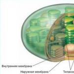 Structure and functions of chloroplasts