Presentation on the topic of the cardiovascular system. Presentation on anatomy on the topic of the cardiovascular system prepared by
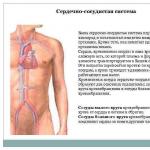
In humans, the heart is located near the center of the chest cavity, it is shifted 2/3 to the left side. The weight of a man's heart is on average 300g, a woman's heart weighs
located near
chest center
cavity, it is 2/3
shifted to the left
side. Heart weight
men are equal in
average 300g,
women - 250g.
flattened in the anteroposterior
direction.
It distinguishes between the top and
base. The apex is the pointed part of the heart,
directed down and to the left and
a little forward. The base is the expanded part of the heart,
facing up and to the right and
a little back. Comprises
durable elastic fabric
heart muscle (myocardium),
which throughout
life rhythmically shortens,
sending blood through the arteries and
capillaries to body tissues.
Structure of the heart
HEART is a powerful muscular organ that pumps bloodthrough a system of cavities (chambers) and valves into a closed
distribution system called system
blood circulation
The wall of the heart consists of
three layers:
internal - endocardium,
middle - myocardium and
external - epicardium. The endocardium lines the inside surface of the chambers of the heart, it
formed by a special type of epithelial tissue - endothelium.
The endothelium has a very smooth, shiny surface that
provides a reduction in friction during blood movement in the heart.
The myocardium makes up the bulk of the heart wall.
It is formed by striated cardiac muscle
fabric, the fibers of which, in turn, are located in
several layers. The atrial myocardium is much thinner than
ventricular myocardium. The myocardium of the left ventricle is three times thicker,
than the myocardium of the right ventricle. Degree of myocardial development
depends on the amount of work performed by the chambers of the heart.
The myocardium of the atria and ventricles is divided by a layer
connective tissue (fibrous ring), which makes it possible
alternate contraction of the atria and ventricles.
The epicardium is a special serous membrane of the heart formed
connective and epithelial tissue.
Chambers of the heart
Heart valves
Jobvalves
hearts
provides
one-sided
movement
blood
in heart.
Blood vessels
representclosed system
hollow elastic
tubes of various
structure, diameter and
mechanical properties.
vessels of the circulatory system
ARTERIESCAPILLARIES
VIENNS
Arteries carry blood from the heart, and veins carry blood
returns to the heart. Between arterial and
venous sections of the circulatory system
the microcirculatory system connecting them is located
bed, including arterioles, venules,
capillaries.
ARTERIES
The artery wall consists of three membranes:internal, middle and external.
The inner lining is the endothelium
(flat epithelium with very smooth
surface).
The middle layer is formed by smooth muscle
tissue and contains well-developed
elastic fibers. Due to smooth
muscle fibers are carried out
change in the lumen of the artery.
Elastic fibers provide
resilience, elasticity and strength
artery walls.
The outer shell consists of loose
fibrous connective tissue,
which plays a protective role and
promotes fixation of arteries in
certain position.
As the arteries move away from the heart, they become stronger
branch, eventually forming the smallest
- arterioles.
CAPILLARIES
The thin wall of capillaries is formed by only onelayer of flat endothelial cells. Through her
blood gases and metabolic products pass easily
substances, nutrients, vitamins, hormones
and leukocytes (if necessary).
Vienna
The structure of the vein wallfundamentally the same as
arteries. But the peculiarity
is significantly smaller
wall thickness due to
subtleties of the middle layer. In him
much less muscle and
elastic fibers due to
low blood pressure in
veins
The second feature of veins is a large number of venous
valves on the inner wall. They are located
in pairs in the form of two semilunar folds. Venous
valves prevent blood from flowing back into
veins during the work of skeletal muscles. Venous
There are no valves in the superior vena cava, in the pulmonary veins,
veins of the brain and heart.
CIRCLES OF BLOOD CIRCULATION
Cardiac cycle.
The sequence of contractions of the chambers of the heart is calledcardiac cycle. During the cycle, each of the four
chambers goes through not only the contraction phase (systole),
but also the relaxation phase (diastole).
The atria contract first: first the right one, almost
immediately behind it is the left one. These cuts provide
rapid filling with blood of the relaxed
ventricles.
Then the ventricles contract, forcefully pushing out
the blood they contain.
At this time, the atria relax and fill
blood from the veins. Each such cycle continues for
on average 6/7 seconds.
Heart work in numbers
In children and adults, the heart contracts at different frequencies: in children under one year old - 100-200 contractions perminute, at 10 years old - 90, and at 20 years old and older - 60-70; after 60 years the number of contractions becomes more frequent and
reaches 90-95. In athletes-runners, during running at sports competitions, the frequency
heart rate can reach up to 250 per minute, the running is over - the heart gradually
calms down, and soon its normal rhythm of contractions is established.
With each contraction, the heart ejects about 60–75 ml of blood, and per minute (at an average frequency
contractions 70 per minute) – 4–5 l. Over 70 years, the heart produces more than 2.5 billion contractions and
pumps approximately 156 million liters of blood.
The work of the heart, like any other work, is measured by the product of the weight of the lifted load (in
kilograms) per height (meters). Let's try to determine its work.
During the day, if a person does not do hard work, the heart contracts over 100,000 times; in a year -
about 40,000,000 times, and over 70 years of life - almost 3,000,000,000 times. What an impressive number - three
billion cuts!
Now multiply the heart rate by the amount of blood ejected, and you will see what
It pumps a huge amount of it. After making the calculation, you will be convinced that in an hour the heart
pumps about 300 liters of blood, per day - over 7000 liters, per year - 2,500,000, and over 70 years of life -
175,000,000 l. The blood that the heart pumps during a person's life can be filled with
4375 railway tanks. If the heart pumped not blood, but water, then from the pumped
In 70 years of water, they could create a lake 2.5 m deep, 7 km wide and 10 km long.
The work of the heart is very significant. So, with one blow, work is done, with the help of which
you can lift a load of 200 g to a height of 1 m. In 1 minute, the heart would lift this load 70 m, i.e.
the height of almost a twenty-story building. If it were possible to use the work of the heart, then in 8 hours
it would be possible to lift a person to the height of the Moscow University building (about 240 m), and in 30-31
day to the top of Chomolungma - the highest point on the globe (8848 m)!
BLOOD PRESSURE
The rhythmic work of the heart creates and maintains the differencepressure in blood vessels. During the contraction of the heart, blood
under pressure is pushed into the arteries. During
blood passing through vessels pressure energy
is wasted. Because blood pressure gradually
decreases. In the aorta it is highest 120-150 mmHg, in
arteries - up to 120 mmHg, in capillaries up to 20, and in hollow
veins from 3-8 mmHg. to the minimum (-5) (below
atmospheric). According to the law of physics, liquid moves from
an area with higher pressure to an area with lower pressure.
Blood pressure is not constant
size. It pulsates in time with the contractions of the heart:
at the moment of systole the pressure rises to 120-130
mmHg. (systolic pressure), and during diastole
decreases to 80-90 mmHg. (diastolic). These
pulse pressure fluctuations occur simultaneously
with pulse fluctuations of the arterial wall.
A person's blood pressure is measured in the brachial
arteries, comparing it with the atmospheric one.
HOW IS BLOOD PRESSURE MEASURED?
The pressure gauge cuff is inflatedair while the pulse is on the wrist
won't disappear. Now shoulder
the artery is compressed by a large
external pressure and blood
it doesn't flow. After,
gradually releasing air from
cuffs, monitor the appearance
pulse At this moment the pressure
there is a little in the artery
greater than the pressure in
cuff, and blood, and with it
and the pulse wave begins
reach the wrist.
Pressure gauge readings in this
time and will characterize
blood pressure in brachial
arteries.
PULSE
Pulse. When contractingventricular blood
ejecting into the aorta,
increasing its pressure.
The wave that arises
while in its wall,
distributed from
certain speed
from the aorta to the arteries.
Rhythmic vibrations
artery walls.
Caused by the rise
pressure in the aorta during
systole is called
pulse.
Pulse can be determined in
places where large arteries
come close to
body surfaces (wrist,
temples, sides of neck).
Presentation ON ANATOMY ON THE TOPIC: CARDIOVASCULAR SYSTEM Prepared by a student of the 21st Sat group of the KRVUZ Crimean Medical College Ibadlaeva Gulnara
 Cardiovascular System Your cardiovascular system transports oxygen and nutrients between tissues and organs. In addition, it helps remove toxins from the body. The heart, blood vessels and blood itself form a complex network through which plasma and formed elements are transported in your body. These substances are carried by the blood through the blood vessels, and the blood drives the heart, which works like a pump. The blood vessels of the cardiovascular system form two main subsystems: the vessels of the pulmonary circulation and the vessels of the systemic circulation. The pulmonary circulation vessels carry blood from the heart to the lungs and back. The vessels of the systemic circulation connect the heart to all other parts of the body.

 Blood vessels carry blood between the heart and various tissues and organs of the body. The following types of blood vessels exist: arteries arterioles capillaries venules and veins Arteries and arterioles carry blood away from the heart. Veins and venules deliver blood back to the heart.
 Arteries and arterioles Arteries carry blood from the ventricles of the heart to other parts of the body. They have a large diameter and thick elastic walls that can withstand very high blood pressure. Before connecting with capillaries, arteries divide into thinner branches called arterioles. Capillaries are the smallest blood vessels that connect arterioles to venules. Due to the very thin wall of the capillaries, they allow the exchange of nutrients and other substances (such as oxygen and carbon dioxide) between the blood and the cells of various tissues. Depending on the need for oxygen and other nutrients, different tissues have different numbers of capillaries. Tissues such as muscles consume large amounts of oxygen and therefore have a dense network of capillaries. On the other hand, tissues with a slow metabolism (such as the epidermis and cornea) do not have capillaries at all. The human body has a lot of capillaries: if they could be unwoven and pulled into one line, then its length would be from 40,000 to 90,000 km!
 Venules and Veins Venules are tiny vessels that connect capillaries to veins, which are larger than venules. Veins run almost parallel to the arteries and carry blood back to the heart. Unlike arteries, veins have thinner walls that contain less muscle and elastic tissue. The Importance of Oxygen The cells of your body need oxygen, and it is the blood that carries oxygen from the lungs to various organs and tissues. When you breathe, oxygen passes through the walls of special air sacs (alveoli) in the lungs and is captured by special blood cells (red blood cells). Oxygen-enriched blood travels through the pulmonary circulation to the heart, which pumps it through the systemic circulation to other parts of the body. Once in different tissues, the blood gives up the oxygen it contains and takes up carbon dioxide instead. Blood saturated with carbon dioxide returns to the heart, which pumps it again to the lungs, where it is freed from carbon dioxide and saturated with oxygen, thereby completing the gas exchange cycle.
 How the Heart Works To pump blood through the heart, its chambers undergo alternating relaxations (diastole) and contractions (systole), during which the chambers fill with blood and push it out accordingly. The right atrium of the heart receives oxygen-poor blood from two main veins: the superior vena cava and the inferior vena cava, as well as from the smaller coronary sinus, which collects blood from the walls of the heart itself. When the right atrium contracts, blood enters the right ventricle through the tricuspid valve. When the right ventricle is sufficiently filled with blood, it contracts and pumps blood through the pulmonary arteries into the pulmonary circulation. Blood enriched with oxygen in the lungs travels through the pulmonary veins to the left atrium. Once filled with blood, the left atrium contracts and pushes blood through the mitral valve into the left ventricle. After filling with blood, the left ventricle contracts and pumps blood into the aorta with great force. From the aorta, blood enters the vessels of the systemic circulation, carrying oxygen to all cells of the body.
Slide 1
The cardiovascular system
The presentation was made by Elena Shakhova, an 8th grade student
Slide 2

The cardiovascular system consists of the circulatory and lymphatic systems. The circulatory system consists of the heart and blood vessels. The vessels that carry blood from the heart to the organs are arteries, and the vessels that bring blood to the heart are veins. The lymphatic system consists of organs of the immune system and lymphatic pathways.
Slide 3

Heart
a hollow muscular organ weighing 240-330 g, cone-shaped, pumping blood into the arteries and receiving venous blood. The heart is located in the chest cavity between the lungs, in the lower mediastinum. has two atria, two ventricles and four valves; receives blood from two vena cava and four pulmonary veins, and throws it into the aorta and pulmonary trunk. The heart pumps 9 liters of blood per day, making from 60 to 160 beats per minute. There are pericardium, myocardium and endocardium. The heart is located in the cardiac sac - the pericardium. Cardiac muscle - the myocardium consists of several layers of muscle fibers; there are more of them in the ventricles than in the atria. These fibers, contracting, push blood from the atria into the ventricles and from the ventricles into the vessels. The internal cavities of the heart and valves are lined by the endocardium.
Slide 4

Inside, the heart is divided by partitions into four chambers. The two atria are divided by the interatrial septum into the left and right atria. The left and right ventricles of the heart are separated by the interventricular septum. Normally, the left and right parts of the heart are completely separate. The atria and ventricles have different functions. The atria store blood that flows into the heart. When the volume of this blood is sufficient, it is pushed into the ventricles. And the ventricles push blood into the arteries, through which it moves throughout the body. The ventricles have to do more hard work, so the muscle layer in the ventricles is much thicker than in the atria. The atria and ventricles on each side of the heart are connected by the atrioventricular orifice. Blood moves through the heart in only one direction. In the large circle of blood circulation from the left part of the heart (left atrium and left ventricle) to the right, and in the small circle from the right to the left. The correct direction is ensured by the valve apparatus of the heart: tricuspid pulmonary mitral aortic valves.
Slide 5

Systemic and pulmonary circulation
The systemic circulation begins in the left ventricle, passes through all internal organs and ends in the right atrium. The pulmonary circulation begins in the right ventricle, passes through the lungs and ends in the left atrium.
Slide 6

Vessels of the systemic circulation
The systemic circulation begins with the largest vessel – the aorta. The aorta is divided into the ascending part, the aortic arch and the descending part. The ascending section begins with a significant expansion - the aortic bulb. The length of this section is about 6 cm. It lies behind the pulmonary trunk and, together with it, is covered by the pericardium. Aortic arch - at the level of the manubrium of the sternum, the aorta bends posteriorly and to the left, spreading over the left main bronchus. The descending section begins at the level of the IV thoracic vertebra. It lies in the posterior mediastinum, at the beginning to the left of the spinal column, gradually deviating to the right, at the level of the XII thoracic vertebra, located anterior to the spine, along the midline. There are two sections of the descending aorta: the thoracic aorta and the abdominal aorta, the division takes place along the aortic notch of the diaphragm. At the level of the IV lumbar vertebra, the descending aorta is divided into its terminal branches - the right and left common iliac arteries, the so-called aortic bifurcation. From the aorta, blood flows through its numerous paired and unpaired branches - arteries - to all parts of the body.
Slide 7

Vessels of the pulmonary circulation
The pulmonary circulation includes: the pulmonary trunk, the right and left pulmonary arteries and their branches, the microcircular bed of the lungs, two right and two left pulmonary veins.
Slide 8

Coronary circle of blood circulation
The coronary circle of blood circulation is cardiac. It includes the vessels of the heart itself to supply blood to the heart muscle. The coronary circle is characterized by the following features: V High pressure, since the coronary vessels begin from the aorta. The coronary vessels form a dense capillary network in the heart muscle with many end-type vessels, which poses a danger if they become blocked, especially in old age. Blood enters the coronary vessels during diastole. This is due to the fact that in the systole phase the mouths of the capillaries are closed by the semilunar valves of the aorta, and also because during systole the myocardium contracts, the coronary vessels are compressed and the flow of blood into them is difficult. During diastole, the myoglobin of the heart muscle is saturated with oxygen, which it very easily gives to the heart in phase. The presence of arteriolovenular anastomoses and arteriolosinusoidal shunts V Special regulation of the tone of the coronary vessels
Slide 9

Arteries
The blood in the arteries is under high pressure. The presence of elastic fibers allows the arteries to pulsate - expand with each heartbeat and collapse when blood pressure drops. Large arteries are divided into medium and small (arterioles), the wall of which has a muscular layer innervated by autonomic vasoconstrictor and vasodilator nerves. The wall of the arteries consists of the inner, middle and outer membranes. The middle shell is separated by an internal elastic membrane from the inner shell and an outer elastic membrane from the outer shell.
Slide 10

Vienna
Having entered the capillaries from the arteries and passed through them, the blood enters the venous system. It first enters very small vessels called venules, which are equivalent to arterioles. The blood continues its journey through small veins and returns to the heart through veins that are large enough to be visible under the skin. These veins contain valves that prevent blood from returning to the tissues. The valves are shaped like a small crescent moon protruding into the lumen of the duct, causing blood to flow in only one direction. Blood enters the venous system, passing through the smallest vessels - capillaries. Exchanges between blood and extracellular fluid occur through the walls of capillaries. Most of the tissue fluid returns to the venous capillaries, and some enters the lymphatic channel. Larger venous vessels can contract or dilate, regulating the flow of blood into them. The movement of the veins is largely due to the tone of the skeletal muscles surrounding the veins, which contract and compress the veins. The pulsation of the arteries adjacent to the veins has a pump effect.
Slide 11

Lymphatic system
The lymphatic system is a part of the vascular system that complements the cardiovascular system. It plays an important role in metabolism and cleansing of body cells and tissues. Unlike the circulatory system, the lymphatic system is not closed and does not have a central pump. The lymph circulating in it moves slowly and under low pressure. The lymphatic system begins in the periphery with “blind” lymphatic capillaries, which become thin lymphatic vessels, which connect into collecting ducts that empty into large veins at the base of the neck. Lymph flowing through the lymphatic vessels is “filtered” in the lymph nodes, which are located along the path of the lymphatic vessels.
Presentation on biology for grade 8 on the topic "Human Cardiovascular System".
1. Composition and significance of the components of the cardiovascular system.
3. Hygiene of the cardiovascular system (hypertension).
Download:
Slide captions:
PRESENTATION HUMAN CARDIOVASCULAR SYSTEM
biology teacher MBOU Mikhailovskaya RV (c) OSHTabakaeva Galina Valentinovna
The cardiovascular system is an organ system that circulates blood in the human body.
1) STRUCTURE OF THE CARDIOVASCULAR SYSTEM
Why is it called "CARDIOVASCULAR"
?!
BECAUSEThe cardiovascular system is formed by:
1. HEART
organ that causes blood to move through blood vessels
hollow tubes of various sizes through which blood circulates.
2. BLOOD VESSELS -
BLOOD VESSELS
ARTERIES
VIENNS
CAPILLARIES
The vessels that carry blood from the heart to the organs are called arteries, and from the organs to the heart - veins.
As blood vessels move away from the heart, they become smaller and form capillaries.
The pulmonary (pulmonary) circulation is limited by blood circulation in the lungs, where the blood is enriched with oxygen and carbon dioxide is removed.
blood circulation provides blood to all organs and tissues
BIG CIRCLE
right ventricle
right atrium
left ventricle
left atrium
inferior vena cava
superior vena cava
pulmonary vein
AORTA
Pulmonary artery
2) BLOOD
BLOOD is the internal environment of the body, formed by liquid connective tissue.
BLOOD TYPES and RH FACTOR:
α and β – first (0); A and β – second (A); α and B – third (B); A and B – fourth (AB).
(Rh +) - Rh-positive group (Rh -) - Rh-negative group
3) CARDIOVASCULAR SYSTEM HYGIENE
HYPERTENSION (HIGH BLOOD PRESSURE)
THE DEVELOPMENT OF HYPERTENSION IS CONTRIBUTED TO: STRESS, PSYCHOEMOTIONAL TENSION; LIFESTYLE (LOW/EXCESSIVE LEVEL OF PHYSICAL ACTIVITY, SEDIENT LIFESTYLE); CONSUMPTION OF LARGE QUANTITIES OF FOOD SALT (NaCl); SMOKING, CONSUMPTION ALCOHOL; DISEASES OF THE ENDOCRINE, URINARY, NERVOUS SYSTEM ORGANS.
RECOMMENDATIONS FOR PREVENTION OF HYPERTENSION
LEAD AN ACTIVE LIFE, TRY TO QUIT SMOKING AND ALCOHOL ABUSE.
MONITOR YOUR BLOOD PRESSURE PERIODICALLY. DO NOT SELF-medicate, SEEK HELP FROM QUALIFIED PROFESSIONALS.
GET YOUR SLEEP! REMEMBER! HEALTHY SLEEP IS THE KEY TO A HEALTHY AND HAPPY LIFE!
TRY NOT TO TAKE EVERYTHING TO YOUR HEART!
THANK YOU FOR YOUR ATTENTION!
On the topic: methodological developments, presentations and notes
A presentation for 8th grade biology students on the topic “Human Urinary System” is designed to help students visualize the structure of the urinary system, its macro and microstructure, as well as...
“Muscle work” - Leg muscles. The structure and function of skeletal muscles. Which letter represents smooth and striated muscles? Physical inactivity. Muscles of the torso at the back. Presentation for 8th grade Protsenko L.V. A-; B-. What is indicated by the numbers 1-; 2-; 3-; 4-. Basic concepts. Independent work: p. 69, Motor unit (MU).
"The Growth of Man" - Judgment Day: Friday, November 13, 2026. Coherence? Possible biological basis of the “Global Crisis”. H. von Foester. …”. I.S. Shklovsky, 1980. N = C / (2025-T) billions, where T is the current time, C is a constant (186 people*years). Nt = 186953/(38 - t). Biological basis of the “Global Crisis”.
“Analyzers” - Studying new material. XI. Temperature. What is the structure of the analyzer? XII. Teaching methods. VIII. Lesson plan. List the analyzers you know. "Brain Tentacles" Tactile.
“Internal environment of the body” - The internal environment of the body has a relative constancy of composition and physicochemical properties. Blood Lymph. The relationship between the components of the internal environment of the body. Tissue fluid. Internal environment of the body Tissue Blood Lymph (intercellular) fluid. Blood Plasma Formed elements: Blood platelets platelets Cells Erythrocytes Leukocytes.
“Bid structure” - Internodes. Opposite (ash, lilac, elderberry). A flower bud is the germ of a reproductive shoot. (Example: elderberry, lilac, willow). Knot. Oak. The structure of a vegetative shoot. Whorled (elodea). Selezneva Alena. Linden. Leaf mosaic. Internal structure of the kidney. Green leaves. Internal structure of a vegetative bud.
“Endocrine glands” - Hormones of the sex glands. ENDOCRINE SYSTEM. Glands of internal and mixed secretion. Thyroid. SIMULATOR 1. Pituitary gland 2. Adrenal glands 3. Thyroid gland 4. Pancreas 5. Sex glands. Municipal educational institution Kazachinskaya secondary school. Lesson plan. Lesson objectives. Insulin Adrenaline Thyroxine Norepinephrine Vasopressin Estradiol Testosterone Endorphin.
We also recommend
 Natural areas of Australia "Lesson presentation" presentation for a geography lesson (grade 7) on the topic
 Presentation on anatomy on the topic of the cardiovascular system prepared by
 Astronomy presentation on the topic "universe"
 Presentation on anatomy on the topic of the cardiovascular system prepared by
 Presentation on the topic: Gravity Universal gravity Presentation on the topic gravity
 Theme of the work “Bionics learning from nature: the latest achievements and the future