Type of tissue, structural features, functions. Plant tissues: structural features and functions
Groups of plant cells with a common function, structure and origin are called plant tissues. The most important of them are: integumentary, basic, excretory, conductive, mechanical and educational. Let's consider the structure and functions of plant tissues.
Educational tissues (meristems)
Located in growth zones:
- on the tops of shoots;
- at the tips of the roots;
- along the stems and roots (cambium or lateral meristem, ensures the growth of stems and roots in thickness).
The meristem cells are actively dividing and do not even have time to grow; they are always young, and therefore do not have vacuoles, their walls are thin, and the nucleus is large.
The activity of the apical meristem of bamboo is striking. It grows literally before our eyes, every hour by 2 - 3 cm!
Integumentary tissues
It is known how quickly peeled fruits dry out, or how easily fruits with broken skins become infected with rot. It is the barrier of the integumentary tissue that ensures the safety of the soft parts of the plant.
There are three types of integumentary tissue:
TOP 4 articleswho are reading along with this
- epidermis;
- periderm;
- crust.
Epidermis (skin)- superficial living cells of various organs. Protects underlying tissues and regulates gas exchange and water evaporation by the plant.

Rice. 1. Epidermal cells under a microscope.
Periderm is formed in woody plants when the green color of the shoot turns brown. The periderm consists of cork cells that protect the shoot from frost, microbes and moisture loss.
Crust- dead tissue. It cannot stretch, following the thickening of the trunk, and cracks.
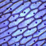
Basic tissues (parenchyma)
There are three types of parenchyma:
- photosynthetic (assimilation);
- aerenchyma, ensures the passage of air into the plant through the intercellular space;
- storing.

Rice. 2. Parenchyma of a green leaf under a microscope.
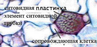
Conductive fabrics
They ensure the movement of substances in the plant body. The movement is carried out in two main directions:
- rising current , carried out by xylem;
- downward current carried out by phloem.
Xylem and phloem form a continuous, plumbing-like system.

Rice. 3. Scheme of the structure of phloem and xylem.
Phloem vessels are composed of sieve elements, or tubes, - elongated cells, the transverse edges of which are similar to a sieve. The flow of substances passes through the pores of the sieve from one cell to another. The cells in the vessel seem to be placed one on one.
The conducting elements of xylem are also represented by elongated cells, but their pores are also located on the side walls of the cells.
Mechanical fabrics
Provide protection and stability of the plant or its individual parts (fruit seeds). Cell membranes are thickened.
Types of mechanical fabric:
- collenchyma (living cells);
- sclerenchyma (dead cells).
Collenchyma is located in growing leaves and stems; it does not interfere with their growth. Contains elongated cells. After the growth of this part of the plant stops, collenchyma gradually turns into sclerenchyma - it becomes tougher, the shells become lignified and thicken.
Lignification increases the fragility of sclerenchyma. Flax fiber is an exception to the rule; it is not lignified sclerenchyma. That's why flax makes such a soft fabric like cambric.
Excretory tissues
These are tissues that secrete water or some secretion from the plant (essential oil, nectar, resin, salts, etc.). This type of tissue also includes those whose secretions remain inside the plant. These are, for example, lacticifers that contain milky juice in their vacuoles (celandine, dandelion).
Their main function is to remove unnecessary substances and protect. Thus, the resin in coniferous wood protects it from rotting.
Using the table “Plant Tissues” we will briefly summarize what has been said:
|
Fabrics |
Functions |
Features of cell structure |
Location |
|
Integumentary |
Protection and gas exchange |
Tight adhesion of cells to each other |
Plant surface |
|
Educational |
Small, with thin walls |
Apical parts of shoots and roots; |
|
|
Mechanical |
Thickened shells |
Stem, leaf veins |
|
|
Basic |
Photosynthesis, food storage. substances |
Loose arrangement of cells |
The basis of the plant, in all organs; stem center |
|
excretory |
Protection and highlighting |
The structure is varied |
Everywhere |
|
Conductive |
Transport of substances |
Vascular elements |
Everywhere |
What have we learned?
From a 6th grade biology paper, we learned that there are six main types of plant tissues. A plant is a system in which tissues are elements. Each tissue provides some area of plant life. Each tissue is vital; the normal development of the entire plant depends on its successful functioning. Tissue cells are specialized; they have structural features corresponding to the functions they perform.
Test on the topic
Evaluation of the report
Average rating: 4.7. Total ratings received: 570.
- Epithelial (integumentary) tissue, or epithelium, is a boundary layer of cells that lines the integument of the body, the mucous membranes of all internal organs and cavities, and also forms the basis of many glands. The epithelium separates the organism (internal environment) from the external environment, but at the same time serves as an intermediary in the interaction of the organism with the environment. Epithelial cells are tightly connected to each other and form a mechanical barrier that prevents the penetration of microorganisms and foreign substances into the body. Epithelial tissue cells live for a short time and are quickly replaced by new ones (this process is called regeneration).
Epithelial tissue is also involved in many other functions: secretion (exocrine and endocrine glands), absorption (intestinal epithelium), gas exchange (lung epithelium).
The main feature of the epithelium is that it consists of a continuous layer of tightly adjacent cells. The epithelium can be in the form of a layer of cells lining all surfaces of the body, and in the form of large accumulations of cells - glands: liver, pancreas, thyroid, salivary glands, etc. In the first case, it lies on the basement membrane, which separates the epithelium from the underlying connective tissue . However, there are exceptions: epithelial cells in the lymphatic tissue alternate with connective tissue elements; such epithelium is called atypical.
Epithelial cells, arranged in a layer, can lie in many layers (stratified epithelium) or in one layer (single-layer epithelium). Based on the height of the cells, epithelia are divided into flat, cubic, prismatic, and cylindrical.
- Connective tissueम will costfrom cells, intercellular substance and connective tissue fibers. It consists of bones, cartilage, tendons, ligaments, blood, fat, it is present in all organs (loose connective tissue) in the form of the so-called stroma (framework) of organs.
In contrast to epithelial tissue, in all types of connective tissue (except adipose tissue), the intercellular substance predominates over the cells in volume, i.e., the intercellular substance is very well expressed. The chemical composition and physical properties of the intercellular substance are very diverse in different types of connective tissue. For example, blood - the cells in it “float” and move freely, since the intercellular substance is well developed.
In general, connective tissue makes up what is called the internal environment of the body. It is very diverse and is represented by various types - from dense and loose forms to blood and lymph, the cells of which are in the liquid. The fundamental differences in the types of connective tissue are determined by the ratios of cellular components and the nature of the intercellular substance.
Dense fibrous connective tissue (muscle tendons, joint ligaments) is dominated by fibrous structures and experiences significant mechanical stress.
Loose fibrous connective tissue is extremely common in the body. It is very rich, on the contrary, in cellular forms of different types. Some of them are involved in the formation of tissue fibers (fibroblasts), others, which is especially important, provide primarily protective and regulatory processes, including through immune mechanisms (macrophages, lymphocytes, tissue basophils, plasma cells).
- Bone.The bone tissue that forms the bones of the skeleton is very strong. It maintains body shape (constitution) and protects organs located in the skull, chest and pelvic cavities, and participates in mineral metabolism. The tissue consists of cells (osteocytes) and intercellular substance in which nutrient channels with blood vessels are located. The intercellular substance contains up to 70% mineral salts (calcium, phosphorus and magnesium).
In its development, bone tissue passes through fibrous and lamellar stages. In various parts of the bone it is organized in the form of compact or spongy bone substance.
- Cartilage tissue. Cartilage tissue consists of cells (chondrocytes) and intercellular substance (cartilage matrix), characterized by increased elasticity. It performs a supporting function, as it forms the main mass of cartilage.
There are three types of cartilage tissue: hyaline , which is part of the cartilage of the trachea, bronchi, ends of the ribs, articular surfaces of bones; elastic , forming the auricle and epiglottis; fibrous , located in the intervertebral discs and joints of the pubic bones.
- Adipose tissue. Adipose tissue is similar to loose connective tissue. The cells are large and filled with fat. Adipose tissue performs nutritional, shape-forming and thermoregulating functions. Adipose tissue is divided into two types: white and brown. In humans, white adipose tissue predominates, part of it surrounds the organs, maintaining their position in the human body and other functions. The amount of brown adipose tissue in humans is small (it is present mainly in newborns). The main function of brown adipose tissue is heat production. Brown adipose tissue maintains the body temperature of animals during hibernation and the temperature of newborn children.
- Muscle.Muscle cells are called muscle fibers because they are constantly stretched in one direction.
Classification of muscle tissue is carried out on the basis of the structure of the tissue (histologically): by the presence or absence of transverse striations, and on the basis of the mechanism of contraction - voluntary (as in skeletal muscle) or involuntary (smooth or cardiac muscle).
Muscle tissue has excitability and the ability to actively contract under the influence of the nervous system and certain substances. Microscopic differences allow us to distinguish two types of this tissue - smooth (unstriated) and striated (striated).
Smooth muscle tissue has a cellular structure. It forms the muscular membranes of the walls of internal organs (intestines, uterus, bladder, etc.), blood and lymphatic vessels; its contraction occurs involuntarily.
Striated muscle tissue consists of muscle fibers, each of which is represented by many thousands of cells, fused, in addition to their nuclei, into one structure. It forms skeletal muscles. We can shorten them at will.
A type of striated muscle tissue is cardiac muscle, which has unique abilities. During life (about 70 years), the heart muscle contracts more than 2.5 million times. No other fabric has such strength potential. Cardiac muscle tissue has transverse striations. However, unlike skeletal muscle, there are special areas where the muscle fibers meet. Thanks to this structure, the contraction of one fiber is quickly transmitted to neighboring ones. This ensures simultaneous contraction of large areas of the heart muscle.
- Nervous tissue.Nervous tissue consists of two types of cells: nerve (neurons) and glial. Glial cells are closely adjacent to the neuron, performing supporting, nutritional, secretory and protective functions.
Neuron is the basic structural and functional unit of nervous tissue. Its main feature is the ability to generate nerve impulses and transmit excitation to other neurons or muscle and glandular cells of working organs. Neurons can consist of a body and processes. Nerve cells are designed to conduct nerve impulses. Having received information on one part of the surface, the neuron very quickly transmits it to another part of its surface. Since the processes of a neuron are very long, information is transmitted over long distances. Most neurons have two types of processes: short, thick, branching near the body - dendrites, and long (up to 1.5 m), thin and branching only at the very end - axons. Axons form nerve fibers.
A nerve impulse is an electrical wave traveling at high speed along a nerve fiber.
Depending on the functions performed and structural features, all nerve cells are divided into three types: sensory, motor (executive) and intercalary. Motor fibers running as part of nerves transmit signals to muscles and glands, sensory fibers transmit information about the state of organs to the central nervous system.
|
Fabric group |
Types of fabrics |
Tissue structure |
Location |
|
| Epithelium | Flat | The surface of the cells is smooth. Cells are tightly adjacent to each other | Skin surface, oral cavity, esophagus, alveoli, nephron capsules | Integumentary, protective, excretory (gas exchange, urine excretion) |
| Glandular | Glandular cells produce secretions | Skin glands, stomach, intestines, endocrine glands, salivary glands | Excretory (secretion of sweat, tears), secretory (formation of saliva, gastric and intestinal juice, hormones) | |
| Ciliated (ciliated) | Consists of cells with numerous hairs (cilia) | Airways | Protective (cilia trap and remove dust particles) | |
| Connective | Dense fibrous | Groups of fibrous, tightly packed cells without intercellular substance | The skin itself, tendons, ligaments, membranes of blood vessels, cornea of the eye | Integumentary, protective, motor |
| Loose fibrous | Loosely arranged fibrous cells intertwined with each other. The intercellular substance is structureless | Subcutaneous fatty tissue, pericardial sac, nervous system pathways | Connects skin to muscles, supports organs in the body, fills gaps between organs. Provides thermoregulation of the body | |
| Cartilaginous | Living round or oval cells lying in capsules, the intercellular substance is dense, elastic, transparent | Intervertebral discs, laryngeal cartilage, trachea, auricle, joint surface | Smoothing the rubbing surfaces of bones. Protection against deformation of the respiratory tract and ears | |
| Bone | Living cells with long processes, interconnected, intercellular substance - inorganic salts and ossein protein | Skeleton bones | Supportive, motor, protective | |
| Blood and lymph | Liquid connective tissue consists of formed elements (cells) and plasma (liquid with organic and mineral substances dissolved in it - serum and fibrinogen protein) | Circulatory system of the whole body | Carries O2 and nutrients throughout the body. Collects CO 2 and dissimilation products. Ensures the constancy of the internal environment, chemical and gas composition of the body. Protective (immunity). Regulatory (humoral) | |
| Muscular | Cross-striped | Multinucleate cylindrical cells up to 10 cm in length, striated with transverse stripes | Skeletal muscles, cardiac muscle | Voluntary movements of the body and its parts, facial expressions, speech. Involuntary contractions (automaticity) of the heart muscle to push blood through the chambers of the heart. Has the properties of excitability and contractility |
| Smooth | Mononuclear cells up to 0.5 mm long with pointed ends | Walls of the digestive tract, blood and lymph vessels, skin muscles | Involuntary contractions of the walls of internal hollow organs. Raising hair on the skin | |
| Nervous | Nerve cells (neurons) | Nerve cell bodies, varied in shape and size, up to 0.1 mm in diameter | Forms the gray matter of the brain and spinal cord | Higher nervous activity. Communication of the organism with the external environment. Centers of conditioned and unconditioned reflexes. Nervous tissue has the properties of excitability and conductivity |
| Short processes of neurons - tree-branching dendrites | Connect with processes of neighboring cells | They transmit the excitation of one neuron to another, establishing a connection between all organs of the body | ||
| Nerve fibers - axons (neurites) - long processes of neurons up to 1.5 m in length. Organs end with branched nerve endings | Nerves of the peripheral nervous system that innervate all organs of the body | Pathways of the nervous system. They transmit excitation from the nerve cell to the periphery via centrifugal neurons; from receptors (innervated organs) - to the nerve cell along centripetal neurons. Interneurons transmit excitation from centripetal (sensitive) neurons to centrifugal (motor) neurons |
Textile it is a collection of cells and intercellular substance that have a common origin, structure and function.
EPITHELIAL TISSUE. Epithelial tissue (epithelium) lines the mucous and serous membranes of internal organs, covers the surfaces of the body and forms numerous glands.
1. Functions:
· separate the internal environment from the external one;
· suction;
· secretion (secretory);
· exchange of substances with the environment;
· protective;
· gas exchange.
2. Structural features and properties:
· cells are located tightly to each other in the form of a layer;
· lie on the border of two environments – external and internal;
There is very little intercellular substance;
layers of cells lie on basement membrane, the nucleus of epithelial cells is shifted to the basal part of the cell;
· there are no blood vessels in the epithelial layers; cell nutrition is carried out by the diffusion of nutrients through the basement membrane;
· rich in nerve fibers and receptors.
· high ability to regenerate.
3. Classification.
Epithelial tissues are divided into:
- single layer squamous epithelium ( mesothelium): lines the surface serous membranes,(peritoneum, pleura, pericardium), forms the wall of the pulmonary alveoli;
- single-layer cubic epithelium forms the walls of the kidney tubules, excretory ducts of the glands, small bronchi;
- single layer columnar epithelium lines the inner surface of the stomach, intestines, uterus, gallbladder, bile ducts and pancreatic duct;
- single-layer multi-row flickering epithelium lines the respiratory tract and some parts of the reproductive system;
- stratified non-keratinizing squamous epithelium lines the cornea of the eye, oral cavity, esophagus;
- stratified keratinizing squamous epithelium lines the surface of the skin;
- transitional epithelium lines the bladder, ureters;
- glandular epithelium forms glands internal(secrets into the internal environment of the body (pituitary gland, adrenal glands)), external(secretes into hollow organs or into the external environment (liver, sweat)) and mixed(secrets into both the external and internal environment (pancreas)) secretions.
CONNECTIVE TISSUE. They are very diverse in structure and functions.
1. Structural features:
· cells are arranged loosely;
There is a lot of intercellular substance;
The intercellular substance contains many fibers ( collagen, elastic, reticular), fills the gaps between cells and fibers basic amorphous substance;
Connective tissue cells are diverse ( fibroblasts, histiocytes, macrophages, mast cells and others).
2. Functions:
unite all the structures of the body into a single whole ( integration);
· mechanical (the basis of organs);
Trophic (participation in metabolism, maintenance homeostasis),
· protective ( phagocytosis and mechanical protection);
· supporting and form-building;
· plastic (participation in regeneration, wound healing).
3. Classification:
The following connective tissues are distinguished in the human body:
- loose fibrous : accompanies blood, lymphatic vessels and nerves, forms the stroma of parenchymal organs; contains a large number of fibers that intertwine in different directions, between them there are cells of different structure and functions;
- dense fibrous : ligaments, tendons, membranes, fascia, membranes of some organs; the fibers are located parallel to each other and form bundles;
- bone : skeletal bones ( lamellar), the intercellular solid substance forms plates in which bone cells are located ( osteocytes, osteoblasts(bone formers), osteoclasts(bone destroyers); if the plates are located at an angle to each other, bone tissue is called spongy; if the plates are located tightly around the bone tubules, the bone tissue is called compact; the structural and functional unit of compact bone tissue is osteon, it is formed by bone plates, which are located in concentric circles around the bone tubule with vessels and nerves; places of attachment of tendons and ligaments ( coarse fiber);
- cartilaginous : auricle, some cartilages of the larynx, including the epiglottis ( elastic cartilage), intervertebral discs, pubic joint, surfaces of the temporomandibular and sternoclavicular joints, places of attachment of ligaments and tendons to bones ( fibrocartilage), most of the articular cartilages, the walls of the airways, the anterior ends of the ribs, the cartilages of the nasal septum ( hyaline cartilage); intercellular substance is dense; There are no blood vessels, and the hyaline cartilage becomes calcified with age.
- reticular : stroma of red bone marrow, lymph nodes, spleen; performs the function of hematopoiesis.
- blood And lymph : part of the internal environment of the body;
- fatty : omentums, subcutaneous fat layer, near organs (for example, kidneys);
- pigmented : near the nipples and anus.
MUSCLE TISSUE. They provide all motor acts in the human body.
1. Main properties:
· excitability;
· conductivity,
· contractility.
2. Structural features:
· have a fibrous structure;
presence of contractile elements myofibrils, represented by proteins, actin And myosin;
· smooth muscle tissues are represented by fusiform, mononuclear cells without transverse striations - myocytes;
· striated are formed by long multinuclear fibers with transverse striations.
3. Functions:
· movement of the body in space, parts of the body relative to each other;
· reduction of internal organs, change in their volume;
· movement of blood through the vessels, food through the gastrointestinal tract, urine, and so on;
· maintaining posture and vertical position of the body in space.
Smooth muscle tissue regenerates well, striated muscle tissue regenerates poorly. Under unfavorable conditions, muscle tissue is replaced by connective tissue, forming a scar.
4. Classification:
- smooth: forms the muscular walls of hollow internal organs (stomach, uterus, bladder, gall bladder and others) and tubular organs (blood vessels, ureters, excretory ducts of glands and others), muscles of the pupil, skin; innervated by fibers of the autonomic nervous system; contracts involuntarily, slowly; gets tired slowly;
- skeletal striated : skeletal muscles, muscles of the mouth, pharynx, partially esophagus; innervated by fibers of the somatic nervous system; contracts voluntarily, quickly; gets tired quickly;
- cardiac striated : heart muscles (myocardium); muscle fibers ( cardiomyocytes) contain one or two nuclei, connected to each other by jumpers, so excitation quickly covers the entire myocardium; innervated by fibers of the autonomic nervous system; contracts involuntarily.
NERVOUS TISSUE. It is the main component of the nervous system. Consists of nerve cells - neurons And neuroglia, playing a supporting role.
1. Main properties:
· excitability;
· conductivity.
2. Functions:
· neurons – generation and conduction of nerve impulses;
· neuroglia in relation to neurons - supporting, trophic, secretory, protective
In the human body it forms all structures of the central and peripheral nervous system.
The structural and functional unit of nervous tissue is the neuron. He has body, which contains the nucleus and all organelles and processes. Numerous short, branching processes are called dendrites, they conduct impulses to the neuron body. Long, unbranched shoot - axon, conducts impulses from the neuron body. Axons are covered with a sheath of fat-like substance - myelin, which has Ranvier interceptions. The sheath acts as an insulator, preventing the dispersion of the nerve impulse.
Based on their functions, neurons are divided into sensitive(conduct impulses to the central nervous system), motor(conduct impulses from the central nervous system to the working organs) and insertion(located between sensitive and motor).
Based on the number of processes, neurons are classified unipolar (pseudounipolar) (one process extends from the body, which branches), bipolar(two processes extend from the body), multipolar (several processes extend from the body).
Tissue as a collection of cells and intercellular substance. Types and types of fabrics, their properties. Intercellular interactions.
There are about 200 types of cells in the adult human body. Groups of cells that have the same or similar structure, are connected by a common origin and are adapted to perform certain functions form fabrics . This is the next level of the hierarchical structure of the human body - the transition from the cellular level to the tissue level (see Figure 1.3.2).
Any tissue is a collection of cells and intercellular substance , which can be a lot (blood, lymph, loose connective tissue) or little (integumentary epithelium).
The cells of each tissue (and some organs) have their own name: the cells of the nervous tissue are called neurons , bone tissue cells - osteocytes , liver - hepatocytes and so on.
Intercellular substance chemically is a system consisting of biopolymers in high concentration and water molecules. It contains structural elements: collagen fibers, elastin, blood and lymph capillaries, nerve fibers and sensory endings (pain, temperature and other receptors). This provides the necessary conditions for the normal functioning of tissues and the performance of their functions.
There are four types of fabrics in total: epithelial , connecting (including blood and lymph), muscular And nervous (see figure 1.5.1).
Epithelial tissue , or epithelium , covers the body, lines the internal surfaces of organs (stomach, intestines, bladder and others) and cavities (abdominal, pleural), and also forms most of the glands. In accordance with this, a distinction is made between the integumentary and glandular epithelium.
Covering epithelium (type A in Figure 1.5.1) forms layers of cells (1), closely - practically without intercellular substance - adjacent to each other. It happens single-layer or multilayer . The integumentary epithelium is a border tissue and performs the main functions: protection from external influences and participation in the metabolism of the body with the environment - absorption of food components and release of metabolic products ( excretion ). The integumentary epithelium is flexible, ensuring the mobility of internal organs (for example, contractions of the heart, distension of the stomach, intestinal motility, expansion of the lungs, and so on).
Glandular epithelium consists of cells, inside of which there are granules with a secret (from the Latin secretio- department). These cells synthesize and secrete many substances important to the body. Through secretion, saliva, gastric and intestinal juices, bile, milk, hormones and other biologically active compounds are formed. The glandular epithelium can form independent organs - glands (for example, the pancreas, thyroid gland, endocrine glands, or endocrine glands , releasing hormones directly into the blood that perform regulatory functions in the body and others), and may be part of other organs (for example, gastric glands).
Connective tissue (types B and C in Figure 1.5.1) is distinguished by a wide variety of cells (1) and an abundance of intercellular substrate, consisting of fibers (2) and amorphous substance (3). Fibrous connective tissue can be loose or dense. Loose connective tissue (type B) is present in all organs, it surrounds blood and lymphatic vessels. Dense connective tissue performs mechanical, supporting, shaping and protective functions. In addition, there is also very dense connective tissue (type B), which consists of tendons and fibrous membranes (dura mater, periosteum, and others). Connective tissue not only performs mechanical functions, but also actively participates in metabolism, the production of immune bodies, the processes of regeneration and wound healing, and ensures adaptation to changing living conditions.
Connective tissue also includes adipose tissue (View D in Figure 1.5.1). Fats are deposited (deposited) in it, the breakdown of which releases a large amount of energy.
Play an important role in the body skeletal (cartilage and bone) connective tissues . They perform mainly supporting, mechanical and protective functions.
Cartilage tissue (type D) consists of cells (1) and a large amount of elastic intercellular substance (2); it forms intervertebral discs, some components of joints, trachea, and bronchi. Cartilage tissue does not have blood vessels and receives the necessary substances by absorbing them from surrounding tissues.
Bone (type E) consists of bone plates, inside of which lie cells. The cells are connected to each other by numerous processes. Bone tissue is hard and the bones of the skeleton are built from this tissue.
A type of connective tissue is blood . In our minds, blood is something very important for the body and, at the same time, difficult to understand. Blood (type G in Figure 1.5.1) consists of intercellular substance - plasma (1) and weighed in it shaped elements (2) - erythrocytes, leukocytes, platelets (Figure 1.5.2 shows their photographs obtained using an electron microscope). All formed elements develop from a common precursor cell. The properties and functions of blood are discussed in more detail in section 1.5.2.3.
Cells muscle tissue (Figure 1.3.1 and types Z and I in Figure 1.5.1) have the ability to contract. Since contraction requires a lot of energy, muscle cells have a higher content mitochondria .
There are two main types of muscle tissue - smooth (type 3 in Figure 1.5.1), which is present in the walls of many, and usually hollow, internal organs (vessels, intestines, gland ducts and others), and striated (view I in Figure 1.5.1), which includes cardiac and skeletal muscle tissue. Bundles of muscle tissue form muscles. They are surrounded by layers of connective tissue and penetrated by nerves, blood and lymphatic vessels (see Figure 1.3.1).
General information on tissues is given in Table 1.5.1.
Table 1.5.1. Tissues, their structure and functions
| Fabric name | Specific cell names | Intercellular substance | Where is this fabric found? | Functions | Drawing |
|---|---|---|---|---|---|
| EPITHELIAL TISSUE | |||||
| Covering epithelium (single-layer and multilayer) | Cells ( epithelial cells ) fit tightly to each other, forming layers. The cells of the ciliated epithelium have cilia, while the cells of the intestinal epithelium have villi. | Small, does not contain blood vessels; the basement membrane demarcates the epithelium from the underlying connective tissue. | The internal surfaces of all hollow organs (stomach, intestines, bladder, bronchi, blood vessels, etc.), cavities (abdominal, pleural, articular), the surface layer of skin ( epidermis ). | Protection from external influences (epidermis, ciliated epithelium), absorption of food components (gastrointestinal tract), excretion of metabolic products (urinary system); ensures organ mobility. | Fig.1.5.1, view A |
| Glandular epithelium |
Glandulocytes contain secretory granules with biologically active substances. They can be located singly or form independent organs (glands). | The intercellular substance of the gland tissue contains blood, lymphatic vessels, and nerve endings. | Glands of internal (thyroid, adrenal glands) or external (salivary, sweat) secretion. Cells can be located singly in the integumentary epithelium (respiratory system, gastrointestinal tract). | Output hormones (section 1.5.2.9), digestive enzymes (bile, gastric, intestinal, pancreatic juice, etc.), milk, saliva, sweat and tear fluid, bronchial secretions, etc. | Rice. 1.5.10 “Skin structure” - sweat and sebaceous glands |
| Connective tissues | |||||
| Loose connective | The cellular composition is characterized by great diversity: fibroblasts , fibrocytes , macrophages , lymphocytes , single adipocytes and etc. | A large number of; consists of an amorphous substance and fibers (elastin, collagen, etc.) | Present in all organs, including muscles, surrounds blood and lymphatic vessels, nerves; main component dermis . | Mechanical (sheath of vessel, nerve, organ); participation in metabolism ( trophism ), the production of immune bodies, processes regeneration . | Fig.1.5.1, view B |
| Dense connecting | Fibers predominate over amorphous matter. | Framework of internal organs, dura mater, periosteum, tendons and ligaments. | Mechanical, shaping, supporting, protective. | Fig.1.5.1, view B | |
| Fat | Almost the entire cytoplasm adipocytes occupies a fat vacuole. | There is more intercellular substance than cells. | Subcutaneous fatty tissue, perinephric tissue, abdominal omentum, etc. | Deposition of fats; energy supply due to the breakdown of fats; mechanical. | Fig.1.5.1, view D |
| Cartilaginous | Chondrocytes , chondroblasts (from lat. chondron- cartilage) | It is distinguished by its elasticity, including due to its chemical composition. | Cartilages of the nose, ears, larynx; articular surfaces of bones; anterior ribs; bronchi, trachea, etc. | Supportive, protective, mechanical. Participates in mineral metabolism (“salt deposition”). Bones contain calcium and phosphorus (almost 98% of the total calcium!). | Fig.1.5.1, view D |
| Bone | Osteoblasts , osteocytes , osteoclasts (from lat. os- bone) | Strength is due to mineral “impregnation”. | Skeletal bones; auditory ossicles in the tympanic cavity (malleus, incus and stapes) | Fig.1.5.1, view E | |
| Blood | Red blood cells (including juvenile forms), leukocytes , lymphocytes , platelets and etc. | Plasma 90-93% consists of water, 7-10% - proteins, salts, glucose, etc. | Internal contents of the cavities of the heart and blood vessels. If their integrity is violated, bleeding and hemorrhage occur. | Gas exchange, participation in humoral regulation, metabolism, thermoregulation, immune defense; coagulation as a defensive reaction. | Fig.1.5.1, view G; Fig.1.5.2 |
| Lymph | Mostly lymphocytes | Plasma (lymphoplasma) | Internal contents of the lymphatic system | Participation in immune defense, metabolism, etc. | Rice. 1.3.4 "Cell Shapes" |
| MUSCLE TISSUE | |||||
| Smooth muscle tissue | Orderly arranged myocytes spindle-shaped | There is little intercellular substance; contains blood and lymphatic vessels, nerve fibers and endings. | In the walls of hollow organs (vessels, stomach, intestines, urinary and gall bladder, etc.) | Peristalsis of the gastrointestinal tract, contraction of the bladder, maintenance of blood pressure due to vascular tone, etc. | Fig.1.5.1, view 3 |
| Cross-striped | Muscle fibers can contain over 100 cores! | Skeletal muscles; cardiac muscle tissue is automatic (chapter 2.6) | Pumping function of the heart; voluntary muscle activity; participation in thermoregulation of the functions of organs and systems. | Fig.1.5.1 (view I) | |
| NERVOUS TISSUE | |||||
| Nervous | Neurons ; neuroglial cells perform auxiliary functions | Neuroglia rich in lipids (fats) | Brain and spinal cord, ganglia (nerve ganglia), nerves (nerve bundles, plexuses, etc.) | Perception of irritation, generation and conduction of impulses, excitability; regulation of the functions of organs and systems. | Fig.1.5.1, view K |
The preservation of shape and the performance of specific functions by the tissue is genetically programmed: the ability to perform specific functions and to differentiate is transmitted to daughter cells via DNA. The regulation of gene expression as the basis of differentiation was discussed in section 1.3.4.
Differentiation is a biochemical process in which relatively homogeneous cells, arising from a common progenitor cell, are transformed into increasingly specialized, specific types of cells that form tissues or organs. Most differentiated cells usually retain their specific characteristics even in a new environment.
In 1952, scientists from the University of Chicago separated chicken embryo cells by growing (incubating) them in an enzyme solution with gentle stirring. However, the cells did not remain separated, but began to unite into new colonies. Moreover, when liver cells mixed with retinal cells, the formation of cellular aggregates occurred in such a way that the retinal cells always moved to the inner part of the cell mass.
Cell interactions . What allows fabrics not to crumble at the slightest external influence? And what ensures the coordinated work of cells and their performance of specific functions?
Many observations prove that cells have the ability to recognize each other and respond accordingly. Interaction is not only the ability to transmit signals from one cell to another, but also the ability to act together, that is, synchronously. On the surface of each cell there are receptors (see section 1.3.2), thanks to which each cell recognizes another similar to itself. And these “detector devices” function according to the “key-lock” rule - this mechanism is repeatedly mentioned in the book.
Let's talk a little about how cells communicate with each other. There are two main methods of intercellular interaction: diffusion And adhesive . Diffusion is an interaction based on intercellular channels, pores in the membranes of neighboring cells located strictly opposite each other. Adhesive (from Latin adhaesio- adhesion, adhesion) - mechanical connection of cells, long-term and stable holding them at a close distance from each other. The chapter on cell structure describes various types of intercellular connections (desmosomes, synapses, and others). This is the basis for the organization of cells into various multicellular structures (tissues, organs).
Each tissue cell not only connects with neighboring cells, but also interacts with the intercellular substance, receiving with its help nutrients, signaling molecules (hormones, mediators), and so on. Through chemicals delivered to all tissues and organs of the body, humoral type of regulation (from Latin humor- liquid).
Another way of regulation, as mentioned above, is carried out using the nervous system. Nerve impulses always reach their target hundreds or thousands of times faster than delivery of chemicals to organs or tissues. The nervous and humoral ways of regulating the functions of organs and systems are closely interrelated. However, the very formation of most chemicals and their release into the blood are under constant control of the nervous system.
Cell, fabric - these are the first levels of organization of living organisms , but even at these stages it is possible to identify general regulatory mechanisms that ensure the vital activity of organs, organ systems and the body as a whole.
In the process of evolution, with the emergence of higher plants onto land, they developed tissues that reached their greatest specialization in flowering plants. In this article we will take a closer look at what plant tissues are, what types they exist, what functions they perform, as well as the structural features of plant tissues.
Fabric are groups of cells that are similar in structure and perform the same functions.
The main plant tissues are shown in the figure below:
Types, functions and structure of plant tissues.


Integumentary tissue of plants.


Plant integumentary tissue - crust
Conductive plant tissue.
| Fabric name | Structure | Location | Functions |
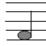
| 1. Wood vessels - xylem | Hollow tubes with lignified walls and dead contents | Wood (xylem) running along the root, stem, leaf veins | Conducting water and minerals from the soil to the root, stem, leaves, flowers |
|
2. Sieve tubes of bast - phloem Accompanying cells or companion cells |
Vertical row of living cells with sieve-like transverse partitions Sister cells of sieve elements that have retained their structure |
Bast (phloem), located along the root, stem, leaf veins Always located along the sieve elements (accompany them) |
Carrying organic matter from leaves to stem, root, flowers Take an active part in carrying organic substances through the sieve tubes of the phloem |
| 3. Conducting vascular-fibrous bundles | A complex of wood and bast in the form of separate strands in grasses and a continuous mass in trees | Central cylinder of root and stem; veins of leaves and flowers | Carrying water and minerals through wood; on bast - organic substances; strengthening organs, connecting them into a single whole |


Mechanical tissue of plants.